Download Mendeley
KAT6A-inhibitor co-crystal structures: tackling a challenging crystallization target via two alternative approaches.
Puetter, V., Bouche, L., Nowak-Reppel, K., Ferrara, S.J., Gradl, S.N., Korr, D., Strathdee, C.A., Ter Laak, A., Hillig, R.C.(2026) Acta Crystallogr D Struct Biol
- PubMed: 42657771 Search on PubMedSearch on PubMed Central
- DOI: https://doi.org/10.1107/S2059798326008545
- Primary Citation Related Structures:
29TM, 29TN, 29UQ, 29UR, 29US, 29UT, 29UU - PubMed Abstract:
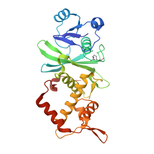
The histone lysine acetyltransferase KAT6A belongs to the MYST family of lysine acetyltransferases which consists of five isoforms. KAT6A (and its paralog KAT6B) have emerged as a promising epigenetic drug targets in cancer, supported by recent clinical data. As support for our high-throughput screening approach aimed at discovering new inhibitors of KAT6A, we developed a crystallization platform system termed KAT6A mutCys in which four surface cysteine residues of KAT6A were mutated to serine. This construct crystallizes readily when in complex with its cofactor AcCoA. The obtained co-crystals were successfully used in a back-soaking approach in which AcCoA was soaked out of the crystals and small-molecule inhibitors were soaked in. In parallel, we explored a previously published surrogate approach termed MYST cryst in which active-site residues of the related, but easier to crystallize, enzyme KAT8 were mutated to those found in KAT6A. By comparing co-crystal structures of the same ligand bound to KAT6A from our Cys-to-Ser approach and to mutated KAT8 (MYST cryst ) we found that the KAT8 surrogate approach indeed successfully reproduced the binding mode observed in the KAT6A structure, while delivering structures with significantly higher resolution. The MYST cryst approach was therefore employed to determine the binding modes of two further small-molecule inhibitors, both from our KAT6A inhibitor optimization program and from a competitor lead series. These structures reveal alternative inhibitor conformations at the binding-site entry and the importance of filling a hydrophobic subpocket in the interior of the binding pocket. These insights will aid future efforts towards the development of KAT6A inhibitors as anticancer drugs.
- Nuvisan ICB GmbH, Muellerstrasse 178, 13353 Berlin, Germany.
Organizational Affiliation: