Download Mendeley
NADPH gating and F-actin engagement define the structural bases of MICAL1 autoinhibition and activation.
Ballabio, F., Minari, A., Bonnet, D.M.V., Graziadei, A., Di Ianni, A., Nobile, M., Tedeschi, G., Camilloni, C., Chaves-Sanjuan, A., Vanoni, M.A.(2026) Arch Biochem Biophys : 111023-111023
- PubMed: 42863552 Search on PubMed
- DOI: https://doi.org/10.1016/j.abb.2026.111023
- Primary Citation Related Structures:
29XK, 29XL - PubMed Abstract:
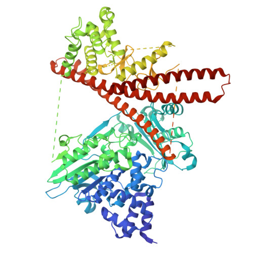
Human MICAL1 is a multidomain protein exhibiting both an oxidase activity, producing H 2 O 2 as a second messenger, and an actin depolymerizing activity through the NADPH-dependent reaction catalyzed by its N-terminal flavoprotein domain. In full-length MICAL1, this domain is inhibited by the following protein regions. Interaction with activating (macro)molecules controls the timing and location of the enzyme activity, explaining its participation in diverse cellular processes. To gain insight into the reaction mechanism and its control, we combined kinetic, cryo-EM, chemical cross-linking and modeling studies of MICAL1, and of complexes with NADPH and F-actin. Contrary to previous models proposing an equilibrium between fully inactive and active conformations, the latter with the properties of the isolated catalytic domain, our results show that MICAL1 exists in solution in a single compact "autoinhibited" conformation able to bind and oxidize NADPH, although at a rate lower than that of the isolated catalytic domain. The reorientation of the gating Trp400 residue is required for productive binding of the nicotinamide portion of NADPH for hydride transfer to the FAD coenzyme, and is specifically hampered in full-length MICAL1 by the Trp400-Phe399-Arg933 triad. In complex with F-actin, the isolated flavoprotein domain bridges two adjacent actin subunits along the filament's ridge with the active site in proximity to, but not in direct contact with, the intersubunit actin interface supporting a mechanism of actin residues oxidation through H 2 O 2 diffusion from the enzyme active site. Model inspection suggests the conformational changes of full-length MICAL1 required for concomitant F-actin binding and relief of "autoinhibition".
- Dipartimento di Bioscienze, Università degli Studi di Milano, Via Celoria 26, 20133 Milano, Italy.
Organizational Affiliation: