2NXX | pdb_00002nxx

Crystal Structure of the Ligand-Binding Domains of the T.castaneum (Coleoptera) Heterodimer EcrUSP Bound to Ponasterone A


Experimental Data Snapshot

  • Method: X-RAY DIFFRACTION
  • Resolution: 2.75 Å
  • R-Value Free: 
    0.281 (Depositor), 0.270 (DCC) 
  • R-Value Work: 
    0.249 (Depositor), 0.240 (DCC) 
  • R-Value Observed: 
    0.249 (Depositor) 

Starting Model: experimental
View more details

wwPDB Validation   3D Report Full Report


Ligand Structure Quality Assessment 


This is version 1.4 of the entry. See complete history


Literature

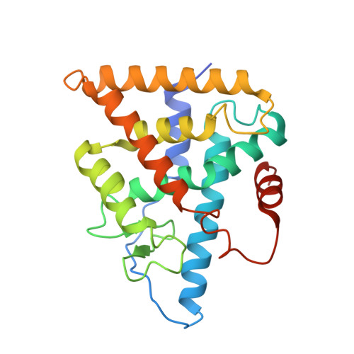
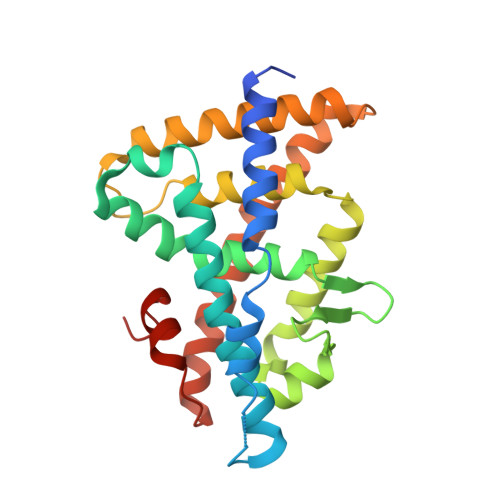
Structural and functional characterization of a novel type of ligand-independent RXR-USP receptor.

Iwema, T.Billas, I.M.Beck, Y.Bonneton, F.Nierengarten, H.Chaumot, A.Richards, G.Laudet, V.Moras, D.

(2007) EMBO J 26: 3770-3782

  • DOI: https://doi.org/10.1038/sj.emboj.7601810
  • Primary Citation of Related Structures:  
    2NXX

  • PubMed Abstract: 

    Retinoid X receptor (RXR) and Ultraspiracle (USP) play a central role as ubiquitous heterodimerization partners of many nuclear receptors. While it has long been accepted that a wide range of ligands can activate vertebrate/mollusc RXRs, the existence and necessity of specific endogenous ligands activating RXR-USP in vivo is still matter of intense debate. Here we report the existence of a novel type of RXR-USP with a ligand-independent functional conformation. Our studies involved Tribolium USP (TcUSP) as representative of most arthropod RXR-USPs, with high sequence homology to vertebrate/mollusc RXRs. The crystal structure of the ligand-binding domain of TcUSP was solved in the context of the functional heterodimer with the ecdysone receptor (EcR). While EcR exhibits a canonical ligand-bound conformation, USP adopts an original apo structure. Our functional data demonstrate that TcUSP is a constitutively silent partner of EcR, and that none of the RXR ligands can bind and activate TcUSP. These findings together with a phylogenetic analysis suggest that RXR-USPs have undergone remarkable functional shifts during evolution and give insight into receptor-ligand binding evolution and dynamics.


  • Organizational Affiliation
    • IGBMC (Institut de Génétique et de Biologie Moléculaire et Cellulaire), UMR7104 CNRS, U596 INSERM, ULP, Département de Biologie et de Génomique Structurales, Illkirch, France.

Macromolecules
Find similar proteins by:  (by identity cutoff)  |  3D Structure
Entity ID: 1
MoleculeChains Sequence LengthOrganismDetailsImage
Ultraspiracle (USP, NR2B4)A,
C [auth B],
E [auth C],
G [auth D]
235Tribolium castaneumMutation(s): 0 
Gene Names: Ultraspiracle (USP)
UniProt
Find proteins for A1JUG2 (Tribolium castaneum)
Explore A1JUG2 
Go to UniProtKB:  A1JUG2
Entity Groups  
Sequence Clusters30% Identity50% Identity70% Identity90% Identity95% Identity100% Identity
UniProt GroupA1JUG2
Sequence Annotations
Expand
  • Reference Sequence
Find similar proteins by:  (by identity cutoff)  |  3D Structure
Entity ID: 2
MoleculeChains Sequence LengthOrganismDetailsImage
Ecdysone Receptor (EcR, NRH1)B [auth E],
D [auth F],
F [auth G],
H
248Tribolium castaneumMutation(s): 0 
Gene Names: Ecdysone Receptor (EcR)
UniProt
Find proteins for A1JUG3 (Tribolium castaneum)
Explore A1JUG3 
Go to UniProtKB:  A1JUG3
Entity Groups  
Sequence Clusters30% Identity50% Identity70% Identity90% Identity95% Identity100% Identity
UniProt GroupA1JUG3
Sequence Annotations
Expand
  • Reference Sequence
Experimental Data & Validation

Experimental Data

  • Method: X-RAY DIFFRACTION
  • Resolution: 2.75 Å
  • R-Value Free:  0.281 (Depositor), 0.270 (DCC) 
  • R-Value Work:  0.249 (Depositor), 0.240 (DCC) 
  • R-Value Observed: 0.249 (Depositor) 
Space Group: P 1 21 1
Unit Cell:
Length ( Å )Angle ( ˚ )
a = 77.54α = 90
b = 89.628β = 91.97
c = 160.247γ = 90
Software Package:
Software NamePurpose
DENZOdata reduction
SCALEPACKdata scaling
CNSrefinement
PDB_EXTRACTdata extraction
ADSCdata collection
HKL-2000data reduction
AMoREphasing

Structure Validation

View Full Validation Report


Ligand Structure Quality Assessment 


Entry History 

Deposition Data

Revision History  (Full details and data files)

  • Version 1.0: 2007-10-02
    Type: Initial release
  • Version 1.1: 2011-07-13
    Changes: Version format compliance
  • Version 1.2: 2017-10-18
    Changes: Refinement description
  • Version 1.3: 2023-12-27
    Changes: Data collection, Database references, Structure summary
  • Version 1.4: 2024-04-03
    Changes: Refinement description