 Download Mendeley
Cryo-EM structure of the L. pneumophila Ceg14 effector in complex with AnkJ
Shatskiy, D., Belyy, A.To be published.
Experimental Data Snapshot
wwPDB Validation 3D Report Full Report

Entity ID: 1 | |||||
|---|---|---|---|---|---|
| Molecule | Chains | Sequence Length | Organism | Details | Image |
| Maltose/maltodextrin-binding periplasmic protein,Dot/Icm secretion system substrate | A [auth B] | 1,060 | Legionella pneumophila | Mutation(s): 0 Gene Names: malE, b4034, JW3994, lpg0437 | 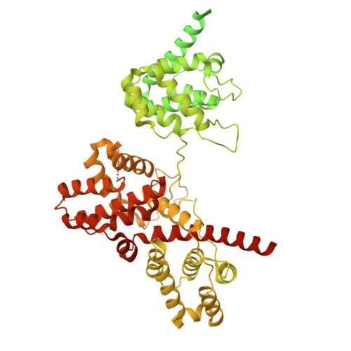 |
UniProt | |||||
Entity Groups | |||||
| Sequence Clusters | 30% Identity50% Identity70% Identity90% Identity95% Identity100% Identity | ||||
| UniProt Groups | P0AEX9Q5ZYD5 | ||||
Sequence AnnotationsExpand | |||||
Reference Sequence | |||||
Entity ID: 2 | |||||
|---|---|---|---|---|---|
| Molecule | Chains | Sequence Length | Organism | Details | Image |
| Ankyrin repeat-containing protein | B [auth C] | 306 | Legionella pneumophila | Mutation(s): 0 Gene Names: legA11, lpg0436 | 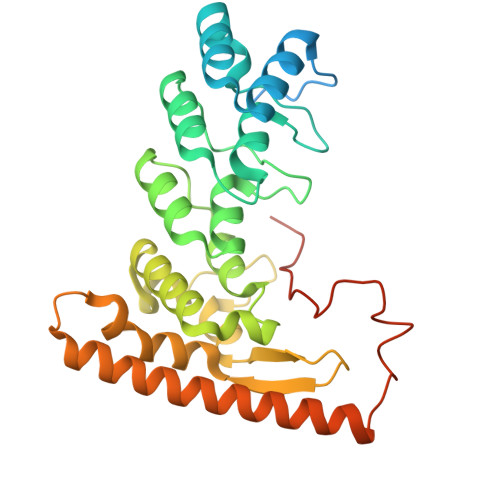 |
UniProt | |||||
Entity Groups | |||||
| Sequence Clusters | 30% Identity50% Identity70% Identity90% Identity95% Identity100% Identity | ||||
| UniProt Group | Q5ZYD6 | ||||
Sequence AnnotationsExpand | |||||
Reference Sequence | |||||
| Task | Software Package | Version |
|---|---|---|
| MODEL REFINEMENT | PHENIX | 1.21.1_5286 |
| RECONSTRUCTION | RELION | 5 |
| Funding Organization | Location | Grant Number |
|---|---|---|
| Netherlands Organisation for Scientific Research (NWO) | Netherlands | OCENW.M.24.262 |
| European Research Council (ERC) | European Union | 101219748 |
