 Download Mendeley
Crystal structure of DDB1 in complex with XS445479
Zeng, H., Ahmad, H., Wang, X., Sun, J., Dong, A., Seitova, A., Edwards, A.M., Arrowsmith, C.H., Peng, H., Halabelian, L., Structural Genomics Consortium (SGC)To be published.
Experimental Data Snapshot
Starting Model: experimental
View more details
wwPDB Validation 3D Report Full Report

Entity ID: 1 | |||||
|---|---|---|---|---|---|
| Molecule | Chains | Sequence Length | Organism | Details | Image |
| DNA damage-binding protein 1 | 1,142 | Homo sapiens | Mutation(s): 0 Gene Names: DDB1, XAP1 | 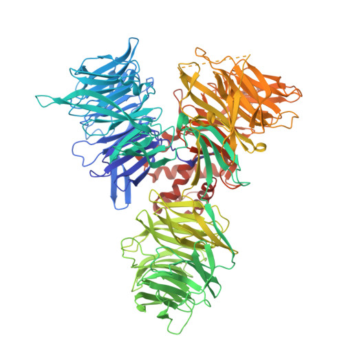 | |
UniProt & NIH Common Fund Data Resources | |||||
PHAROS: Q16531 GTEx: ENSG00000167986 | |||||
Entity Groups | |||||
| Sequence Clusters | 30% Identity50% Identity70% Identity90% Identity95% Identity100% Identity | ||||
| UniProt Group | Q16531 | ||||
Sequence AnnotationsExpand | |||||
Reference Sequence | |||||
Entity ID: 2 | |||||
|---|---|---|---|---|---|
| Molecule | Chains | Sequence Length | Organism | Details | Image |
| UNK | 4 | Homo sapiens | Mutation(s): 0 |  | |
Sequence AnnotationsExpand | |||||
Reference Sequence | |||||
| Ligands 4 Unique | |||||
|---|---|---|---|---|---|
| ID | Chains | Name / Formula / InChI Key | 2D Diagram | 3D Interactions | |
| A1DPN( Subject of Investigation/LOI) Download:Ideal Coordinates CCD File | H [auth A] | 1-[(5-benzyl-1,3,4-oxadiazol-2-yl)methyl]-3-chloro-5-(trifluoromethyl)pyridin-2(1H)-one C16 H11 Cl F3 N3 O2 ARQWSAPXBXDNPF-UHFFFAOYSA-N |  | ||
| TLA Download:Ideal Coordinates CCD File | G [auth A], I [auth A], J [auth A], K [auth A] | L(+)-TARTARIC ACID C4 H6 O6 FEWJPZIEWOKRBE-JCYAYHJZSA-N | 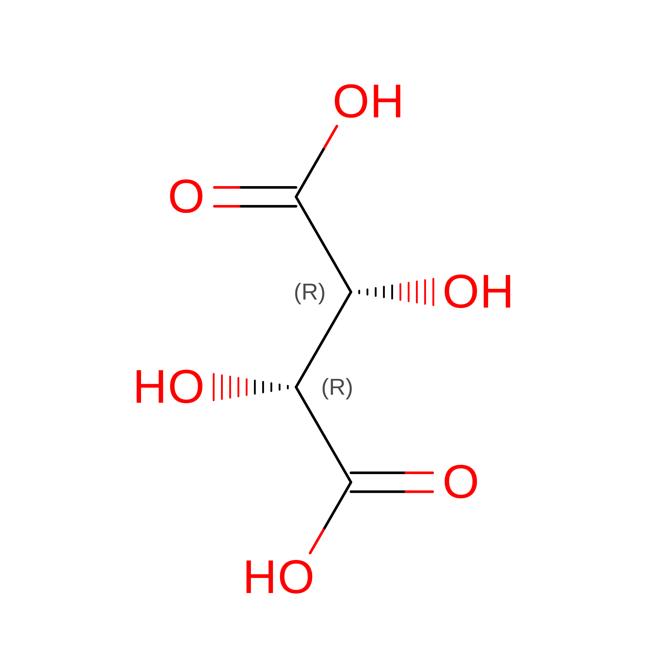 | ||
| EDO Download:Ideal Coordinates CCD File | C [auth A], D [auth A], E [auth A], F [auth A] | 1,2-ETHANEDIOL C2 H6 O2 LYCAIKOWRPUZTN-UHFFFAOYSA-N |  | ||
| UNX Download:Ideal Coordinates CCD File | L [auth A], M [auth A] | UNKNOWN ATOM OR ION X |  | ||
| Modified Residues 1 Unique | |||||
|---|---|---|---|---|---|
| ID | Chains | Type | Formula | 2D Diagram | Parent |
| UNK Query on UNK | B | L-PEPTIDE LINKING | C4 H9 N O2 | 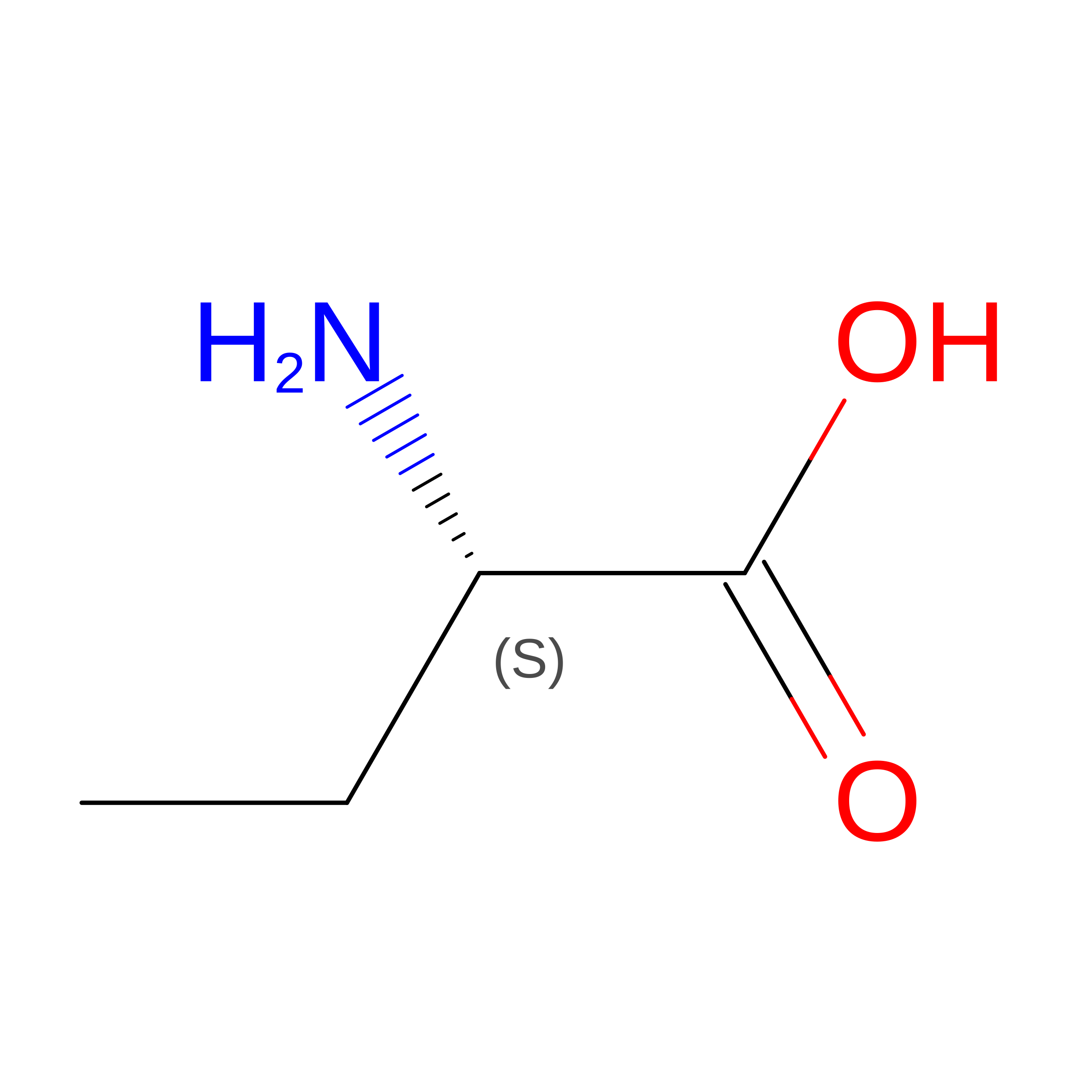 | -- |
| Length ( Å ) | Angle ( ˚ ) |
|---|---|
| a = 62.544 | α = 90 |
| b = 124.709 | β = 90 |
| c = 168.627 | γ = 90 |
| Software Name | Purpose |
|---|---|
| REFMAC | refinement |
| HKL-3000 | data scaling |
| HKL-3000 | data reduction |
| REFMAC | phasing |
| PDB_EXTRACT | data extraction |
| Funding Organization | Location | Grant Number |
|---|---|---|
| Other private | Canada | -- |
