4CHG | pdb_00004chg

Crystal structure of VapBC15 complex from Mycobacterium tuberculosis


Experimental Data Snapshot

  • Method: X-RAY DIFFRACTION
  • Resolution: 2.10 Å
  • R-Value Free: 
    0.200 (Depositor), 0.195 (DCC) 
  • R-Value Work: 
    0.167 (Depositor), 0.165 (DCC) 
  • R-Value Observed: 
    0.169 (Depositor) 

wwPDB Validation   3D Report Full Report


This is version 1.3 of the entry. See complete history


Literature

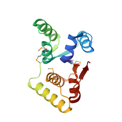
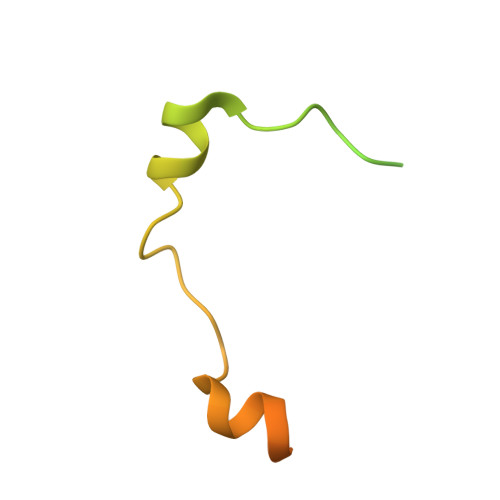
Crystal Structure of the Vapbc-15 Complex from Mycobacterium Tuberculosis Reveals a Two-Metal Ion Dependent Pin-Domain Ribonuclease And a Variable Mode of Toxin-Antitoxin Assembly.

Das, U.Pogenberg, V.Subhramanyam, U.K.T.Wilmanns, M.Gourinath, S.Srinivasan, A.

(2014) J Struct Biol 188: 249

  • DOI: https://doi.org/10.1016/j.jsb.2014.10.002
  • Primary Citation of Related Structures:  
    4CHG

  • PubMed Abstract: 

    Although PIN (PilT N-terminal)-domain proteins are known to have ribonuclease activity, their specific mechanism of action remains unknown. VapCs form a family of ribonucleases that possess a PIN-domain assembly and are known as toxins. The activities of VapCs are impaired by VapB antitoxins. Here we present the crystal structure of the VapBC-15 toxin-antitoxin complex from Mycobacterium tuberculosis determined to 2.1Å resolution. The VapB-15 and VapC-15 components assemble into one heterotetramer (VapB2C2) and two heterotrimers (VapBC2) in each asymmetric unit of the crystal. The active site of VapC-15 toxin consists of a cluster of acidic amino acid residues and two divalent metal ions, forming a well organised ribonuclease active site. The distribution of the catalytic-site residues of the VapC-15 toxin is similar to that of T4 RNase H and of Methanococcus jannaschii FEN-1, providing strong evidence that these three proteins share a similar mechanism of activity. The presence of both VapB2C2 and VapBC2 emphasizes the fact that the same antitoxin can bind the toxin in 1:1 and 1:2 ratios. The crystal structure determination of the VapBC-15 complex reveals for the first time a PIN-domain ribonuclease protein that shows two metal ions at the active site and a variable mode of toxin-antitoxin assembly. The structure further shows that VapB-15 antitoxin binds to the same groove meant for the binding of putative substrate (RNA), resulting in the inhibition of VapC-15's toxicity.


  • Organizational Affiliation
    • Department of Biophysics, All India Institute of Medical Sciences, New Delhi, India.

Macromolecules
Find similar proteins by:  (by identity cutoff)  |  3D Structure
Entity ID: 1
MoleculeChains Sequence LengthOrganismDetailsImage
PROBABLE RIBONUCLEASE VAPC15
A, B, C, D, E
A, B, C, D, E, F
133Mycobacterium tuberculosis H37RvMutation(s): 0 
EC: 3.1
UniProt
Find proteins for P9WF97 (Mycobacterium tuberculosis (strain ATCC 25618 / H37Rv))
Explore P9WF97 
Go to UniProtKB:  P9WF97
Entity Groups  
Sequence Clusters30% Identity50% Identity70% Identity90% Identity95% Identity100% Identity
UniProt GroupP9WF97
Sequence Annotations
Expand
  • Reference Sequence
Find similar proteins by:  (by identity cutoff)  |  3D Structure
Entity ID: 2
MoleculeChains Sequence LengthOrganismDetailsImage
ANTITOXIN VAPB15
G, H, I, J
88Mycobacterium tuberculosis H37RvMutation(s): 0 
EC: 3.1
UniProt
Find proteins for P9WLM7 (Mycobacterium tuberculosis (strain ATCC 25618 / H37Rv))
Explore P9WLM7 
Go to UniProtKB:  P9WLM7
Entity Groups  
Sequence Clusters30% Identity50% Identity70% Identity90% Identity95% Identity100% Identity
UniProt GroupP9WLM7
Sequence Annotations
Expand
  • Reference Sequence
Small Molecules
Ligands 2 Unique
IDChains Name / Formula / InChI Key2D Diagram3D Interactions
MN
Query on MN

Download Ideal Coordinates CCD File 
K [auth A],
M [auth B],
O [auth D],
Q [auth F]
MANGANESE (II) ION
Mn
WAEMQWOKJMHJLA-UHFFFAOYSA-N
MG
Query on MG

Download Ideal Coordinates CCD File 
L [auth A],
N [auth B],
P [auth D],
R [auth F]
MAGNESIUM ION
Mg
JLVVSXFLKOJNIY-UHFFFAOYSA-N
Modified Residues  1 Unique
IDChains TypeFormula2D DiagramParent
MSE
Query on MSE
A, B, C, D, E
A, B, C, D, E, F
L-PEPTIDE LINKINGC5 H11 N O2 SeMET
Experimental Data & Validation

Experimental Data

  • Method: X-RAY DIFFRACTION
  • Resolution: 2.10 Å
  • R-Value Free:  0.200 (Depositor), 0.195 (DCC) 
  • R-Value Work:  0.167 (Depositor), 0.165 (DCC) 
  • R-Value Observed: 0.169 (Depositor) 
Space Group: P 21 21 21
Unit Cell:
Length ( Å )Angle ( ˚ )
a = 86.16α = 90
b = 131.2β = 90
c = 149.35γ = 90
Software Package:
Software NamePurpose
PHENIXrefinement
iMOSFLMdata reduction
SCALEPACKdata scaling
Auto-Rickshawphasing

Structure Validation

View Full Validation Report


Entry History 

Deposition Data

Revision History  (Full details and data files)

  • Version 1.0: 2014-11-12
    Type: Initial release
  • Version 1.1: 2014-12-17
    Changes: Data collection, Database references
  • Version 1.2: 2018-03-07
    Changes: Data collection, Source and taxonomy
  • Version 1.3: 2024-10-23
    Changes: Data collection, Database references, Derived calculations, Other, Structure summary