5JLG | pdb_00005jlg

The X-ray structure of the adduct formed in the reaction between bovine pancreatic ribonuclease and compound I, a piano-stool organometallic Ru(II) arene compound containing an O,S-chelating ligand


Experimental Data Snapshot

  • Method: X-RAY DIFFRACTION
  • Resolution: 1.79 Å
  • R-Value Free: 
    0.236 (Depositor), 0.230 (DCC) 
  • R-Value Work: 
    0.172 (Depositor), 0.180 (DCC) 
  • R-Value Observed: 
    0.175 (Depositor) 

Starting Model: experimental
View more details

wwPDB Validation   3D Report Full Report


Ligand Structure Quality Assessment 


This is version 1.3 of the entry. See complete history


Literature

Unusual mode of protein binding by a cytotoxic pi-arene ruthenium(ii) piano-stool compound containing an O,S-chelating ligand.

Hildebrandt, J.Gorls, H.Hafner, N.Ferraro, G.Durst, M.Runnebaum, I.B.Weigand, W.Merlino, A.

(2016) Dalton Trans 45: 12283-12287

  • DOI: https://doi.org/10.1039/c6dt02380k
  • Primary Citation of Related Structures:  
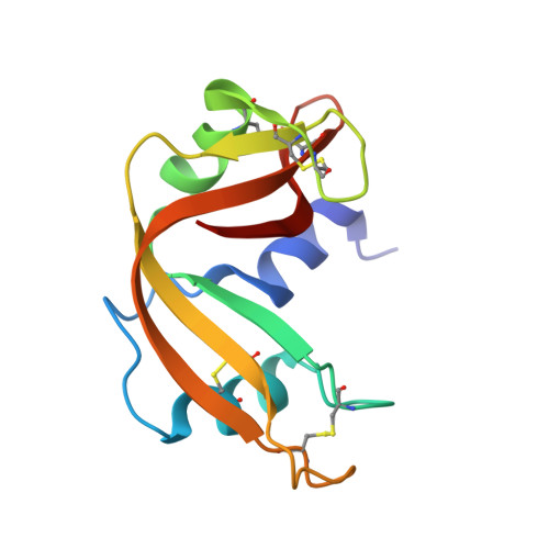
    5JLG

  • PubMed Abstract: 

    A new pseudo-octahedral π-arene ruthenium(ii) piano-stool compound, containing an O,S-bidentate ligand (compound 1) and showing significant cytotoxic activity in vitro, was synthesized and characterized. In solution stability and interaction with the model protein bovine pancreatic ribonuclease (RNase A) were investigated by using UV-Vis absorption spectroscopy. Its crystal structure and that of the adduct formed upon reaction with RNase A were obtained by X-ray crystallography. The comparison between the structure of purified compound 1 and that of the fragment bound to RNase A reveals an unusual mode of protein binding that includes ligand exchange and alteration of coordination sphere geometry.


  • Organizational Affiliation
    • Department of Inorganic and Analytical Chemistry, University of Jena, Germany. wolfgang.weigand@uni-jena.de.

Macromolecules
Find similar proteins by:  (by identity cutoff)  |  3D Structure
Entity ID: 1
MoleculeChains Sequence LengthOrganismDetailsImage
Ribonuclease pancreatic
A, B
124Bos taurusMutation(s): 0 
Gene Names: RNASE1RNS1
EC: 3.1.27.5 (PDB Primary Data), 4.6.1.18 (UniProt)
UniProt
Find proteins for P61823 (Bos taurus)
Explore P61823 
Go to UniProtKB:  P61823
Entity Groups  
Sequence Clusters30% Identity50% Identity70% Identity90% Identity95% Identity100% Identity
UniProt GroupP61823
Sequence Annotations
Expand
  • Reference Sequence
Small Molecules
Ligands 3 Unique
IDChains Name / Formula / InChI Key2D Diagram3D Interactions
6L6
Query on 6L6

Download Ideal Coordinates CCD File 
D [auth A],
G [auth B]
methyl (2Z)-3-hydroxy-3-(3-hydroxyphenyl)prop-2-ene(dithioate)
C10 H10 O2 S2
MOMLKGNSDNGKFJ-TWGQIWQCSA-N
RU
Query on RU

Download Ideal Coordinates CCD File 
C [auth A],
F [auth B]
RUTHENIUM ION
Ru
BPEVHDGLPIIAGH-UHFFFAOYSA-N
DMS
Query on DMS

Download Ideal Coordinates CCD File 
E [auth A],
H [auth B],
I [auth B]
DIMETHYL SULFOXIDE
C2 H6 O S
IAZDPXIOMUYVGZ-UHFFFAOYSA-N
Experimental Data & Validation

Experimental Data

  • Method: X-RAY DIFFRACTION
  • Resolution: 1.79 Å
  • R-Value Free:  0.236 (Depositor), 0.230 (DCC) 
  • R-Value Work:  0.172 (Depositor), 0.180 (DCC) 
  • R-Value Observed: 0.175 (Depositor) 
Space Group: C 1 2 1
Unit Cell:
Length ( Å )Angle ( ˚ )
a = 100.51α = 90
b = 32.7β = 90.03
c = 72.47γ = 90
Software Package:
Software NamePurpose
REFMACrefinement
HKL-2000data reduction
HKL-2000data scaling
PHASERphasing

Structure Validation

View Full Validation Report


Ligand Structure Quality Assessment 


Entry History 

Deposition Data

Revision History  (Full details and data files)

  • Version 1.0: 2016-08-03
    Type: Initial release
  • Version 1.1: 2016-08-10
    Changes: Database references
  • Version 1.2: 2024-01-10
    Changes: Data collection, Database references, Derived calculations, Refinement description
  • Version 1.3: 2024-10-23
    Changes: Structure summary