Structural basis for the specific cleavage of core-fucosylatedN-glycans by endo-beta-N-acetylglucosaminidase from the fungusCordyceps militaris.
Seki, H., Huang, Y., Arakawa, T., Yamada, C., Kinoshita, T., Iwamoto, S., Higuchi, Y., Takegawa, K., Fushinobu, S.(2019) J Biological Chem 294: 17143-17154
- PubMed: 31548313
- DOI: https://doi.org/10.1074/jbc.RA119.010842
- Primary Citation of Related Structures:
6KPL, 6KPM, 6KPN, 6KPO - PubMed Abstract:
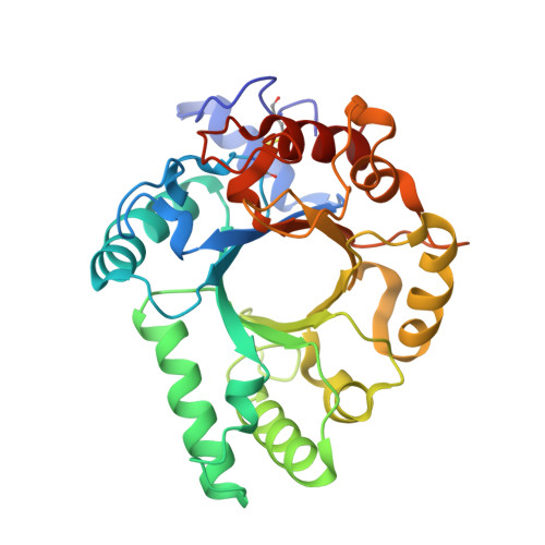
N -Linked glycans play important roles in various cellular and immunological events. Endo-β- N -acetylglucosaminidase (ENGase) can release or transglycosylate N -glycans and is a promising tool for the chemoenzymatic synthesis of glycoproteins with homogeneously modified glycans. The ability of ENGases to act on core-fucosylated glycans is a key factor determining their therapeutic utility because mammalian N -glycans are frequently α-1,6-fucosylated. Although the biochemistries and structures of various ENGases have been studied extensively, the structural basis for the recognition of the core fucose and the asparagine-linked GlcNAc is unclear. Herein, we determined the crystal structures of a core fucose-specific ENGase from the caterpillar fungus Cordyceps militaris (Endo-CoM), which belongs to glycoside hydrolase family 18. Structures complexed with fucose-containing ligands were determined at 1.75-2.35 Å resolutions. The fucose moiety linked to GlcNAc is extensively recognized by protein residues in a round-shaped pocket, whereas the asparagine moiety linked to the GlcNAc is exposed to the solvent. The N -glycan-binding cleft of Endo-CoM is Y-shaped, and several lysine and arginine residues are present at its terminal regions. These structural features were consistent with the activity of Endo-CoM on fucose-containing glycans on rituximab (IgG) and its preference for a sialobiantennary substrate. Comparisons with other ENGases provided structural insights into their core fucose tolerance and specificity. In particular, Endo-F3, a known core fucose-specific ENGase, has a similar fucose-binding pocket, but the surrounding residues are not shared with Endo-CoM. Our study provides a foothold for protein engineering to develop enzymatic tools for the preparation of more effective therapeutic antibodies.
- Department of Biotechnology, University of Tokyo, 1-1-1 Yayoi, Bunkyo-ku, Tokyo 113-8657, Japan.
Organizational Affiliation: