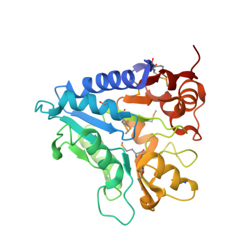
6NBG | pdb_00006nbg

2.05 Angstrom Resolution Crystal Structure of Hypothetical Protein KP1_5497 from Klebsiella pneumoniae.


Experimental Data Snapshot

  • Method: X-RAY DIFFRACTION
  • Resolution: 2.05 Å
  • R-Value Free: 
    0.231 (Depositor), 0.240 (DCC) 
  • R-Value Work: 
    0.201 (Depositor), 0.210 (DCC) 
  • R-Value Observed: 
    0.202 (Depositor) 

wwPDB Validation   3D Report Full Report


This is version 1.2 of the entry. See complete history


Literature

A Structural Systems Biology Approach to High-Risk CG23 Klebsiella pneumoniae.

Inniss, N.L.Kochan, T.J.Minasov, G.Wawrzak, Z.Chang, C.Tan, K.Shuvalova, L.Kiryukhina, O.Pshenychnyi, S.Wu, R.Dubrovska, I.Babnigg, G.Endres, M.Anderson, W.F.Hauser, A.R.Joachimiak, A.Satchell, K.J.F.

(2023) Microbiol Resour Announc 12: e0101322-e0101322

  • DOI: https://doi.org/10.1128/mra.01013-22
  • Primary Citation of Related Structures:  
    6DT3, 6DUX, 6DVV, 6DXN, 6E85, 6NAU, 6NBG, 6NDI, 6WN5, 6WN8, 6X1L, 7RJJ, 7TL5, 7TZP

  • PubMed Abstract: 

    Klebsiella pneumoniae is a leading cause of antibiotic-resistant-associated deaths in the world. Here, we report the deposition of 14 structures of enzymes from both the core and accessory genomes of sequence type 23 (ST23) K1 hypervirulent K. pneumoniae.


  • Organizational Affiliation
    • Department of Microbiology-Immunology, Feinberg School of Medicine, Northwestern University, Chicago, Illinois, USA.

Macromolecules
Find similar proteins by:  (by identity cutoff)  |  3D Structure
Entity ID: 1
MoleculeChains Sequence LengthOrganismDetailsImage
Glucosamine-6-phosphate deaminase
A, B, C, D, E
A, B, C, D, E, F
242Klebsiella pneumoniaeMutation(s): 0 
Gene Names: nagB_2DM059_30720SAMEA4364603_04465
EC: 3.5.99.6
UniProt
Find proteins for A0A332QZQ2 (Klebsiella pneumoniae)
Explore A0A332QZQ2 
Go to UniProtKB:  A0A332QZQ2
Entity Groups  
Sequence Clusters30% Identity50% Identity70% Identity90% Identity95% Identity100% Identity
UniProt GroupA0A332QZQ2
Sequence Annotations
Expand
  • Reference Sequence
Small Molecules
Ligands 2 Unique
IDChains Name / Formula / InChI Key2D Diagram3D Interactions
PO4
Query on PO4

Download Ideal Coordinates CCD File 
G [auth A]
H [auth B]
J [auth D]
K [auth D]
L [auth E]
G [auth A],
H [auth B],
J [auth D],
K [auth D],
L [auth E],
M [auth E],
N [auth F],
O [auth F]
PHOSPHATE ION
O4 P
NBIIXXVUZAFLBC-UHFFFAOYSA-K
CL
Query on CL

Download Ideal Coordinates CCD File 
I [auth C]CHLORIDE ION
Cl
VEXZGXHMUGYJMC-UHFFFAOYSA-M
Modified Residues  1 Unique
IDChains TypeFormula2D DiagramParent
MSE
Query on MSE
A, B, C, D, E
A, B, C, D, E, F
L-PEPTIDE LINKINGC5 H11 N O2 SeMET
Experimental Data & Validation

Experimental Data

  • Method: X-RAY DIFFRACTION
  • Resolution: 2.05 Å
  • R-Value Free:  0.231 (Depositor), 0.240 (DCC) 
  • R-Value Work:  0.201 (Depositor), 0.210 (DCC) 
  • R-Value Observed: 0.202 (Depositor) 
Space Group: C 2 2 21
Unit Cell:
Length ( Å )Angle ( ˚ )
a = 75.482α = 90
b = 123.806β = 90
c = 299.664γ = 90
Software Package:
Software NamePurpose
REFMACrefinement
HKL-3000data reduction
HKL-3000data scaling
HKL-3000phasing

Structure Validation

View Full Validation Report


Entry History 

Revision History  (Full details and data files)

  • Version 1.0: 2018-12-19
    Type: Initial release
  • Version 1.1: 2023-06-14
    Changes: Database references, Refinement description
  • Version 1.2: 2024-10-09
    Changes: Data collection, Structure summary