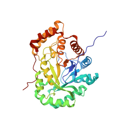
7EZI | pdb_00007ezi

Rice L-galactose dehydrogenase (apo form)


Experimental Data Snapshot

  • Method: X-RAY DIFFRACTION
  • Resolution: 1.20 Å
  • R-Value Free: 
    0.187 (Depositor), 0.205 (DCC) 
  • R-Value Work: 
    0.152 (Depositor) 

Starting Model: experimental
View more details

wwPDB Validation 3D Report Full Report

Validation slider image for 7EZI

This is version 1.2 of the entry. See complete history. 

Literature

Crystal structure of rice L-galactose dehydrogenase.

Fujimoto, Z., Momma, M., Suzuki, N.

(2026) Acta Crystallogr F Struct Biol Commun 82: 289-294

  • DOI: https://doi.org/10.1107/S2053230X26007430
  • Primary Citation Related Structures: 
    7EZI, 7EZL

  • PubMed Abstract: 

    L-Galactose dehydrogenase plays a key role in the biosynthesis of L-ascorbic acid (vitamin C) in plants. Here, we report crystal structures of rice (Oryza sativa) L-galactose dehydrogenase in both apo and NAD + -bound forms at 1.2 and 1.8 Å resolution, respectively. Rice L-galactose dehydrogenase adopts a conserved (β/α) 8 -barrel fold with a well preserved NAD + -binding cleft, as in other plant L-galactose dehydrogenases. Structural comparisons revealed conformational flexibility in the first and second loop regions, which form the lid and side wall of the NAD + -binding site. These loops undergo significant rearrangement upon NAD + binding, likely facilitating cofactor and substrate uptake. These findings suggest a flexible loop-mediated regulation of cofactor and substrate access.


  • Organizational Affiliation: 
    • Research Center for Advanced Analysis, National Agriculture and Food Research Organization, 2-1-2 Kannondai, Tsukuba, Ibaraki 305-8518, Japan.

Macromolecule Content 

  • Total Structure Weight: 35.32 kDa 
  • Atom Count: 2,770 
  • Modeled Residue Count: 323 
  • Deposited Residue Count: 324 
  • Unique protein chains: 1

Macromolecules

Find similar proteins by:|  3D Structure
Entity ID: 1
MoleculeChains  Sequence LengthOrganismDetailsImage
L-galactose dehydrogenase324Oryza sativa Japonica GroupMutation(s): 0 
UniProt
Find proteins for Q2QQV2 (Oryza sativa subsp. japonica)
Explore Q2QQV2 
Go to UniProtKB:  Q2QQV2
Entity Groups
Sequence Clusters30% Identity50% Identity70% Identity90% Identity95% Identity100% Identity
UniProt GroupQ2QQV2
Sequence Annotations
Expand
Reference Sequence

Small Molecules

Ligands 1 Unique
IDChains Name / Formula / InChI Key2D Diagram3D Interactions
MG

Query on MG


Download:Ideal Coordinates CCD File
B [auth A]MAGNESIUM ION
Mg
JLVVSXFLKOJNIY-UHFFFAOYSA-N

Experimental Data & Validation

Experimental Data

  • Method: X-RAY DIFFRACTION
  • Resolution: 1.20 Å
  • R-Value Free:  0.187 (Depositor), 0.205 (DCC) 
  • R-Value Work:  0.152 (Depositor) 
Space Group: P 1 21 1
Unit Cell:
Length ( Å )Angle ( ˚ )
a = 46.745α = 90
b = 54.877β = 102.268
c = 56.753γ = 90
Software Package:
Software NamePurpose
REFMACrefinement
HKL-2000data reduction
SCALEPACKdata scaling
MOLREPphasing

Structure Validation

View Full Validation Report


Entry History 

Deposition Data

Revision History  (Full details and data files)

  • Version 1.0: 2021-06-23
    Type: Initial release
  • Version 1.1: 2023-11-29
    Changes: Data collection, Database references, Derived calculations, Refinement description
  • Version 1.2: 2026-10-28
    Changes: Database references, Structure summary