8K6R | pdb_00008k6r

LnaB-Actin-PRUb ternary complex in the presence of AMPPNP


Experimental Data Snapshot

  • Method: X-RAY DIFFRACTION
  • Resolution: 2.76 Å
  • R-Value Free: 
    0.261 (Depositor), 0.247 (DCC) 
  • R-Value Work: 
    0.205 (Depositor), 0.209 (DCC) 
  • R-Value Observed: 
    0.209 (Depositor) 

Starting Model: experimental
View more details

wwPDB Validation   3D Report Full Report


Ligand Structure Quality Assessment 


This is version 1.1 of the entry. See complete history


Literature

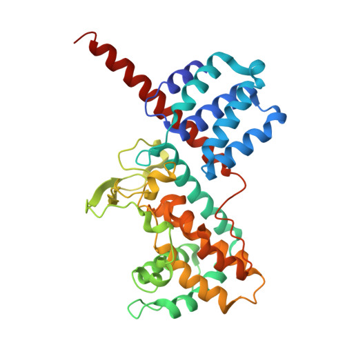
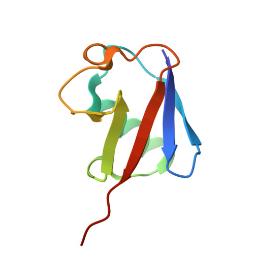
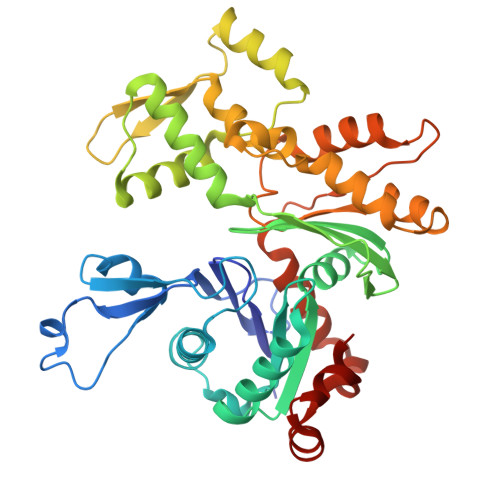
Structure and mechanism of an actin-dependent bacterial phosphoryl AMPylase.

Chen, T.T.Lu, Q.Zheng, S.R.Fu, J.Chen, J.Kang, L.Wu, J.Luo, J.Tong, J.Li, S.Li, X.Li, S.Li, J.Wang, S.Feng, Y.Luo, Z.Q.Ouyang, S.

(2025) Nat Chem Biol 

  • DOI: https://doi.org/10.1038/s41589-025-01945-w
  • Primary Citation of Related Structures:  
    8K6F, 8K6I, 8K6R, 8K6V

  • PubMed Abstract: 

    The two effectors LnaB and MavL of Legionella pneumophila coordinate the conversion of phosphoribosyl ubiquitin (PR-Ub) released by reversal of ubiquitination induced by members of the SidE effector family into functional Ub. LnaB acts as an actin-dependent phosphoryl AMPylase that converts PR-Ub into ADP-ribosylated (ADPR)-Ub. Catalysis by LnaB requires the conserved SHE motif present in a large family of bacterial toxins. Here we describe a series of structures of LnaB in complex with the cofactor actin and the substrate PR-Ub and ATP. LnaB harbors both adenylyltransferase and ATPase activities, which reveal an adenylylation mechanism involved in a two-step catalytic process. Actin performs a unique activation mechanism that promotes the recruitment of PR-Ub by LnaB to activate LnaB's ATPase activity through interacting with LnaB and PR-Ub. Mechanisms derived from this series of structures covering the process of LnaB action establish an important biochemical basis for protein AMPylation.


  • Organizational Affiliation

    Key Laboratory of Microbial Pathogenesis and Interventions of Fujian Province University, Key Laboratory of Innate Immune Biology of Fujian Province, Biomedical Research Center of South China, College of Life Sciences, Fujian Normal University, Fuzhou, China.


Macromolecules
Find similar proteins by:  (by identity cutoff)  |  3D Structure
Entity ID: 1
MoleculeChains Sequence LengthOrganismDetailsImage
LnaB352LegionellaMutation(s): 0 
Entity Groups  
Sequence Clusters30% Identity50% Identity70% Identity90% Identity95% Identity100% Identity
Sequence Annotations
Expand
  • Reference Sequence
Find similar proteins by:  (by identity cutoff)  |  3D Structure
Entity ID: 2
MoleculeChains Sequence LengthOrganismDetailsImage
UbiquitinB [auth C]76Homo sapiensMutation(s): 0 
Gene Names: UBB
UniProt & NIH Common Fund Data Resources
Find proteins for P0CG48 (Homo sapiens)
Explore P0CG48 
Go to UniProtKB:  P0CG48
PHAROS:  P0CG48
GTEx:  ENSG00000150991 
Entity Groups  
Sequence Clusters30% Identity50% Identity70% Identity90% Identity95% Identity100% Identity
UniProt GroupP0CG48
Sequence Annotations
Expand
  • Reference Sequence
Find similar proteins by:  (by identity cutoff)  |  3D Structure
Entity ID: 3
MoleculeChains Sequence LengthOrganismDetailsImage
Actin gamma 1C [auth B]375Oryctolagus cuniculusMutation(s): 0 
UniProt
Find proteins for A0A8C6VAB1 (Naja naja)
Explore A0A8C6VAB1 
Go to UniProtKB:  A0A8C6VAB1
Entity Groups  
Sequence Clusters30% Identity50% Identity70% Identity90% Identity95% Identity100% Identity
UniProt GroupA0A8C6VAB1
Sequence Annotations
Expand
  • Reference Sequence
Small Molecules
Ligands 2 Unique
IDChains Name / Formula / InChI Key2D Diagram3D Interactions
AR6 (Subject of Investigation/LOI)
Query on AR6

Download Ideal Coordinates CCD File 
E [auth A][(2R,3S,4R,5R)-5-(6-AMINOPURIN-9-YL)-3,4-DIHYDROXY-OXOLAN-2-YL]METHYL[HYDROXY-[[(2R,3S,4R,5S)-3,4,5-TRIHYDROXYOXOLAN-2-YL]METHOXY]PHOSPHORYL] HYDROGEN PHOSPHATE
C15 H23 N5 O14 P2
SRNWOUGRCWSEMX-ZQSHOCFMSA-N
ANP (Subject of Investigation/LOI)
Query on ANP

Download Ideal Coordinates CCD File 
D [auth A]PHOSPHOAMINOPHOSPHONIC ACID-ADENYLATE ESTER
C10 H17 N6 O12 P3
PVKSNHVPLWYQGJ-KQYNXXCUSA-N
Experimental Data & Validation

Experimental Data

  • Method: X-RAY DIFFRACTION
  • Resolution: 2.76 Å
  • R-Value Free:  0.261 (Depositor), 0.247 (DCC) 
  • R-Value Work:  0.205 (Depositor), 0.209 (DCC) 
  • R-Value Observed: 0.209 (Depositor) 
Space Group: P 21 21 2
Unit Cell:
Length ( Å )Angle ( ˚ )
a = 217.97α = 90
b = 55.903β = 90
c = 80.458γ = 90
Software Package:
Software NamePurpose
PHENIXrefinement
HKL-2000data reduction
autoPROCdata scaling
PHASERphasing

Structure Validation

View Full Validation Report


Ligand Structure Quality Assessment 


Entry History & Funding Information

Deposition Data


Funding OrganizationLocationGrant Number
National Natural Science Foundation of China (NSFC)China--

Revision History  (Full details and data files)

  • Version 1.0: 2025-01-29
    Type: Initial release
  • Version 1.1: 2025-08-13
    Changes: Database references, Derived calculations, Structure summary