8P3C | pdb_00008p3c

Full length structure of BpMIP with bound inhibitor NJS227.


Experimental Data Snapshot

  • Method: X-RAY DIFFRACTION
  • Resolution: 2.02 Å
  • R-Value Free: 
    0.235 (Depositor), 0.250 (DCC) 
  • R-Value Work: 
    0.194 (Depositor), 0.220 (DCC) 
  • R-Value Observed: 
    0.196 (Depositor) 

Starting Model: experimental
View more details

wwPDB Validation   3D Report Full Report


Ligand Structure Quality Assessment 


This is version 1.2 of the entry. See complete history


Literature

Structure and Dynamics of Macrophage Infectivity Potentiator Proteins from Pathogenic Bacteria and Protozoans Bound to Fluorinated Pipecolic Acid Inhibitors.

Perez Carrillo, V.H.Whittaker, J.J.Wiedemann, C.Harder, J.M.Lohr, T.Jamithireddy, A.K.Dajka, M.Goretzki, B.Joseph, B.Guskov, A.Harmer, N.J.Holzgrabe, U.Hellmich, U.A.

(2025) J Med Chem 68: 5926-5941

  • DOI: https://doi.org/10.1021/acs.jmedchem.5c00134
  • Primary Citation of Related Structures:  
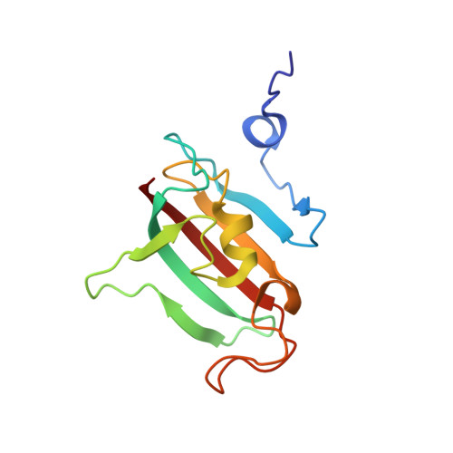
    8P3C, 8P3D, 8P42

  • PubMed Abstract: 

    Macrophage infectivity potentiator (MIP) proteins, found in pro- and eukaryotic pathogens, influence microbial virulence, host cell infection, pathogen replication, and dissemination. MIPs share an FKBP (FK506 binding protein)-like prolyl- cis/trans -isomerase domain, making them attractive targets for inhibitor development. We determined high-resolution crystal structures of Burkholderia pseudomallei and Trypanosoma cruzi MIPs in complex with fluorinated pipecolic acid inhibitors. The inhibitor binding profiles in solution were compared across B. pseudomallei , T. cruzi , and Legionella pneumophila MIPs using 1 H, 15 N, and 19 F NMR spectroscopy. Demonstrating the versatility of fluorinated ligands for characterizing inhibitor complexes, 19 F NMR spectroscopy identified differences in ligand binding dynamics across MIPs. EPR spectroscopy and SAXS further revealed inhibitor-induced global structural changes in homodimeric L. pneumophila MIP. This study demonstrates the importance of integrating diverse methods to probe protein dynamics and provides a foundation for optimizing MIP-targeted inhibitors in this structurally conserved yet dynamically variable protein family.


  • Organizational Affiliation
    • Faculty of Chemistry and Earth Sciences, Institute of Organic Chemistry and Macromolecular Chemistry, Friedrich Schiller University Jena, 07743 Jena, Germany.

Macromolecules
Find similar proteins by:  (by identity cutoff)  |  3D Structure
Entity ID: 1
MoleculeChains Sequence LengthOrganismDetailsImage
Peptidyl-prolyl cis-trans isomeraseA [auth B]126Burkholderia pseudomalleiMutation(s): 0 
Gene Names: fbpBPSS1823
EC: 5.2.1.8
UniProt
Find proteins for Q63J95 (Burkholderia pseudomallei (strain K96243))
Explore Q63J95 
Go to UniProtKB:  Q63J95
Entity Groups  
Sequence Clusters30% Identity50% Identity70% Identity90% Identity95% Identity100% Identity
UniProt GroupQ63J95
Sequence Annotations
Expand
  • Reference Sequence
Small Molecules
Experimental Data & Validation

Experimental Data

  • Method: X-RAY DIFFRACTION
  • Resolution: 2.02 Å
  • R-Value Free:  0.235 (Depositor), 0.250 (DCC) 
  • R-Value Work:  0.194 (Depositor), 0.220 (DCC) 
  • R-Value Observed: 0.196 (Depositor) 
Space Group: P 31 2 1
Unit Cell:
Length ( Å )Angle ( ˚ )
a = 77.608α = 90
b = 77.608β = 90
c = 52.995γ = 120
Software Package:
Software NamePurpose
REFMACrefinement
XDSdata scaling
XDSdata reduction
PHASERphasing

Structure Validation

View Full Validation Report


Ligand Structure Quality Assessment 


Entry History & Funding Information

Deposition Data


Funding OrganizationLocationGrant Number
German Research Foundation (DFG)Germany390713860

Revision History  (Full details and data files)

  • Version 1.0: 2024-06-12
    Type: Initial release
  • Version 1.1: 2025-03-05
    Changes: Database references, Structure summary
  • Version 1.2: 2025-03-26
    Changes: Database references