8SRV | pdb_00008srv

Crystal structure of O-acetyl-L-serine sulfhydrylase A (CysK) from Staphylococcus aureus NCTC 8325 complexed with a transcriptional repressor (CymR) derived 10 amino acid peptide


Experimental Data Snapshot

  • Method: X-RAY DIFFRACTION
  • Resolution: 1.32 Å
  • R-Value Free: 
    0.187 (Depositor), 0.188 (DCC) 
  • R-Value Work: 
    0.164 (Depositor), 0.165 (DCC) 
  • R-Value Observed: 
    0.166 (Depositor) 

Starting Model: experimental
View more details

wwPDB Validation   3D Report Full Report


This is version 1.1 of the entry. See complete history


Literature

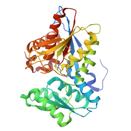
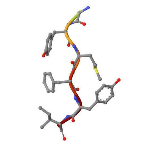
Identification of Cysteine Metabolism Regulator (CymR)-Derived Pentapeptides as Nanomolar Inhibitors of Staphylococcus aureus O -Acetyl-l-serine Sulfhydrylase (CysK).

Pederick, J.L.Vandborg, B.C.George, A.Bovermann, H.Boyd, J.M.Freundlich, J.S.Bruning, J.B.

(2025) ACS Infect Dis 11: 238-248

  • DOI: https://doi.org/10.1021/acsinfecdis.4c00832
  • Primary Citation of Related Structures:  
    8SRT, 8SRU, 8SRV, 8SRW, 8T2C

  • PubMed Abstract: 

    The pathway of bacterial cysteine biosynthesis is gaining traction for the development of antibiotic adjuvants. Bacterial cysteine biosynthesis is generally facilitated by two enzymes possessing O -acetyl-l-serine sulfhydrylases (OASS), CysK and CysM. In Staphylococcus aureus , there exists a single OASS homologue, Sa CysK. Knockout of Sa CysK was found to increase sensitivity to oxidative stress, making it a relevant target for inhibitor development. Sa CysK also forms two functional complexes via interaction with the preceding enzyme in the pathway serine acetyltransferase (CysE) or the transcriptional regulator of cysteine metabolism (CymR). These interactions occur through insertion of a C-terminal peptide of CysE or CymR into the active site of Sa CysK, inhibiting OASS activity, and therefore represent an excellent starting point for developing Sa CysK inhibitors. Here, we detail the characterization of CysE and CymR-derived C-terminal peptides as inhibitors of Sa CysK. Using a combination of X-ray crystallography, surface plasmon resonance, and enzyme inhibition assays, it was determined that the CymR-derived decapeptide forms extensive interactions with Sa CysK and acts as a potent inhibitor ( K D = 25 nM; IC 50 = 180 nM), making it a promising lead for the development of Sa CysK inhibitors. To understand the determinants of this high-affinity interaction, the structure-activity relationships of 16 rationally designed peptides were also investigated. This identified that the C-terminal pentapeptide of CymR facilitates the high-affinity interaction with Sa CysK and that subtle structural modification of the pentapeptide is possible without impacting potency. Ultimately, this work identified CymR pentapeptides as a promising scaffold for the development of antibiotic adjuvants targeting Sa CysK.


  • Organizational Affiliation
    • Institute for Photonics and Advanced Sensing (IPAS), School of Biological Sciences, The University of Adelaide, Adelaide, South Australia 5005, Australia.

Macromolecules
Find similar proteins by:  (by identity cutoff)  |  3D Structure
Entity ID: 1
MoleculeChains Sequence LengthOrganismDetailsImage
Cysteine synthase
A, B
318Staphylococcus aureus subsp. aureus NCTC 8325Mutation(s): 0 
Gene Names: SAOUHSC_00488
EC: 2.5.1.47
UniProt
Find proteins for Q2G0Q8 (Staphylococcus aureus (strain NCTC 8325 / PS 47))
Explore Q2G0Q8 
Go to UniProtKB:  Q2G0Q8
Entity Groups  
Sequence Clusters30% Identity50% Identity70% Identity90% Identity95% Identity100% Identity
UniProt GroupQ2G0Q8
Sequence Annotations
Expand
  • Reference Sequence

Find similar proteins by:  Sequence   |   3D Structure  

Entity ID: 2
MoleculeChains Sequence LengthOrganismDetailsImage
Transcriptional repressor (CymR) derived 10 amino acid peptide
C, D
10Staphylococcus aureus subsp. aureus NCTC 8325Mutation(s): 0 
Entity Groups  
Sequence Clusters30% Identity50% Identity70% Identity90% Identity95% Identity100% Identity
Sequence Annotations
Expand
  • Reference Sequence
Small Molecules
Modified Residues  1 Unique
IDChains TypeFormula2D DiagramParent
LLP
Query on LLP
A, B
L-PEPTIDE LINKINGC14 H22 N3 O7 PLYS
Experimental Data & Validation

Experimental Data

  • Method: X-RAY DIFFRACTION
  • Resolution: 1.32 Å
  • R-Value Free:  0.187 (Depositor), 0.188 (DCC) 
  • R-Value Work:  0.164 (Depositor), 0.165 (DCC) 
  • R-Value Observed: 0.166 (Depositor) 
Space Group: P 1 21 1
Unit Cell:
Length ( Å )Angle ( ˚ )
a = 56.395α = 90
b = 96.24β = 111.92
c = 58.994γ = 90
Software Package:
Software NamePurpose
XDSdata reduction
Aimlessdata scaling
PHASERphasing
PHENIXrefinement
PDB_EXTRACTdata extraction

Structure Validation

View Full Validation Report


Entry History & Funding Information

Deposition Data


Funding OrganizationLocationGrant Number
Not funded--

Revision History  (Full details and data files)

  • Version 1.0: 2024-11-13
    Type: Initial release
  • Version 1.1: 2025-05-28
    Changes: Database references