8T0W | pdb_00008t0w

Crystal structure of dimethylsulfone (DMSO2) monooxygenase SfnG from Pseudomonas fluorescens with DMSO2 and oxidized FMN bound


Experimental Data Snapshot

  • Method: X-RAY DIFFRACTION
  • Resolution: 1.75 Å
  • R-Value Free: 
    0.182 (Depositor), 0.183 (DCC) 
  • R-Value Work: 
    0.155 (Depositor), 0.158 (DCC) 
  • R-Value Observed: 
    0.157 (Depositor) 

Starting Model: experimental
View more details

wwPDB Validation   3D Report Full Report


Ligand Structure Quality Assessment 


This is version 1.0 of the entry. See complete history


Literature

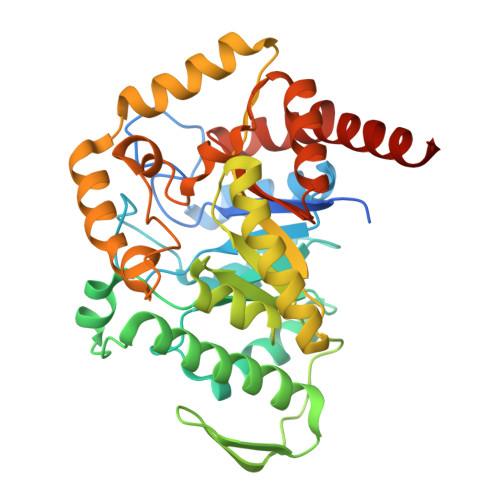
Structural, biophysical, and biochemical insights into C-S bond cleavage by dimethylsulfone monooxygenase.

Gonzalez, R.Soule, J.Phan, N.Wicht, D.K.Dowling, D.P.

(2024) Proc Natl Acad Sci U S A 121: e2401858121-e2401858121

  • DOI: https://doi.org/10.1073/pnas.2401858121
  • Primary Citation of Related Structures:  
    8T0U, 8T0W

  • PubMed Abstract: 

    Sulfur is an essential element for life. Bacteria can obtain sulfur from inorganic sulfate; but in the sulfur starvation-induced response, Pseudomonads employ two-component flavin-dependent monooxygenases (TC-FMOs) from the msu and sfn operons to assimilate sulfur from environmental compounds including alkanesulfonates and dialkylsulfones. Here, we report binding studies of oxidized FMN to enzymes involved within the P. fluorescens enzymatic pathway responsible for converting dimethylsulfone (DMSO 2 ) to sulfite. In this catabolic pathway, SfnG serves as the initial TC-FMO for sulfur assimilation, which is investigated in detail by solving the 2.6-Å resolution crystal structure of unliganded SfnG and the 1.75-Å resolution crystal structure of the SfnG ternary complex containing FMN and DMSO 2 . We find that SfnG adopts a (β/α) 8 barrel fold with a distinct quaternary configuration from other tetrameric class C TC-FMOs. To probe the unexpected tetramer arrangement, structural heterogeneity is assessed by chromatography and light scattering to confirm ligand binding correlates with a tetramer. Binding of FMN and DMSO 2 accompanies ordering of the active site, with DMSO 2 bound on the si -face of the flavin. A previously unobserved protein backbone conformation is found within the oxygen-binding site on the re -face of the flavin. Functional assays and the positioning of ligands with respect to the oxygen-binding site are consistent with use of an N5-(hydro)peroxyflavin pathway. Biochemical endpoint assays and docking studies reveal SfnG breaks the C-S bond of a range of dialkylsulfones.


  • Organizational Affiliation
    • Department of Chemistry, University of Massachusetts, Boston, MA 02125.

Macromolecules
Find similar proteins by:  (by identity cutoff)  |  3D Structure
Entity ID: 1
MoleculeChains Sequence LengthOrganismDetailsImage
FMNH(2)-dependent dimethylsulfone monooxygenase
A, B, C, D, E
A, B, C, D, E, F, G, H
387Pseudomonas fluorescens Pf0-1Mutation(s): 0 
Gene Names: sfnGPfl01_2879
EC: 1.14.14.35
UniProt
Find proteins for Q3KC85 (Pseudomonas fluorescens (strain Pf0-1))
Explore Q3KC85 
Go to UniProtKB:  Q3KC85
Entity Groups  
Sequence Clusters30% Identity50% Identity70% Identity90% Identity95% Identity100% Identity
UniProt GroupQ3KC85
Sequence Annotations
Expand
  • Reference Sequence
Small Molecules
Ligands 4 Unique
IDChains Name / Formula / InChI Key2D Diagram3D Interactions
FMN (Subject of Investigation/LOI)
Query on FMN

Download Ideal Coordinates CCD File 
BA [auth F]
EA [auth G]
HA [auth H]
I [auth A]
M [auth B]
BA [auth F],
EA [auth G],
HA [auth H],
I [auth A],
M [auth B],
Q [auth C],
U [auth D],
Y [auth E]
FLAVIN MONONUCLEOTIDE
C17 H21 N4 O9 P
FVTCRASFADXXNN-SCRDCRAPSA-N
XZ5 (Subject of Investigation/LOI)
Query on XZ5

Download Ideal Coordinates CCD File 
CA [auth F]
FA [auth G]
IA [auth H]
J [auth A]
N [auth B]
CA [auth F],
FA [auth G],
IA [auth H],
J [auth A],
N [auth B],
R [auth C],
V [auth D],
Z [auth E]
(methanesulfonyl)methane
C2 H6 O2 S
HHVIBTZHLRERCL-UHFFFAOYSA-N
GOL
Query on GOL

Download Ideal Coordinates CCD File 
K [auth A],
O [auth B],
S [auth C],
W [auth D]
GLYCEROL
C3 H8 O3
PEDCQBHIVMGVHV-UHFFFAOYSA-N
CL
Query on CL

Download Ideal Coordinates CCD File 
AA [auth E]
DA [auth F]
GA [auth G]
JA [auth H]
L [auth A]
AA [auth E],
DA [auth F],
GA [auth G],
JA [auth H],
L [auth A],
P [auth B],
T [auth C],
X [auth D]
CHLORIDE ION
Cl
VEXZGXHMUGYJMC-UHFFFAOYSA-M
Experimental Data & Validation

Experimental Data

  • Method: X-RAY DIFFRACTION
  • Resolution: 1.75 Å
  • R-Value Free:  0.182 (Depositor), 0.183 (DCC) 
  • R-Value Work:  0.155 (Depositor), 0.158 (DCC) 
  • R-Value Observed: 0.157 (Depositor) 
Space Group: C 2 2 21
Unit Cell:
Length ( Å )Angle ( ˚ )
a = 104.198α = 90
b = 172.727β = 90
c = 397.356γ = 90
Software Package:
Software NamePurpose
PHENIXrefinement
XDSdata reduction
XDSdata scaling
PHENIXphasing

Structure Validation

View Full Validation Report


Ligand Structure Quality Assessment 


Entry History & Funding Information

Deposition Data


Funding OrganizationLocationGrant Number
National Science Foundation (NSF, United States)United States1807480

Revision History  (Full details and data files)

  • Version 1.0: 2024-11-27
    Type: Initial release