8VLB | pdb_00008vlb

Crystal structure of EloBC-VHL-CDO1 complex bound to compound 4 molecular glue


Experimental Data Snapshot

  • Method: X-RAY DIFFRACTION
  • Resolution: 2.90 Å
  • R-Value Free: 
    0.258 (Depositor), 0.260 (DCC) 
  • R-Value Work: 
    0.208 (Depositor), 0.210 (DCC) 
  • R-Value Observed: 
    0.210 (Depositor) 

Starting Model: experimental
View more details

wwPDB Validation   3D Report Full Report


Ligand Structure Quality Assessment 


This is version 1.1 of the entry. See complete history


Literature

A small-molecule VHL molecular glue degrader for cysteine dioxygenase 1.

Tutter, A.Buckley, D.Golosov, A.A.Ma, X.Shu, W.McKay, D.J.J.Darsigny, V.Dovala, D.Beckwith, R.Solomon, J.Rao, P.Xu, L.Fazal, A.Lingel, A.Wartchow, C.Cobb, J.S.Hachey, A.Tullai, J.Michaud, G.A.

(2025) Nat Chem Biol 

  • DOI: https://doi.org/10.1038/s41589-025-01936-x
  • Primary Citation of Related Structures:  
    8VL9, 8VLB

  • PubMed Abstract: 

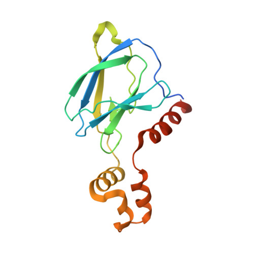
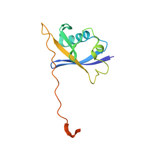
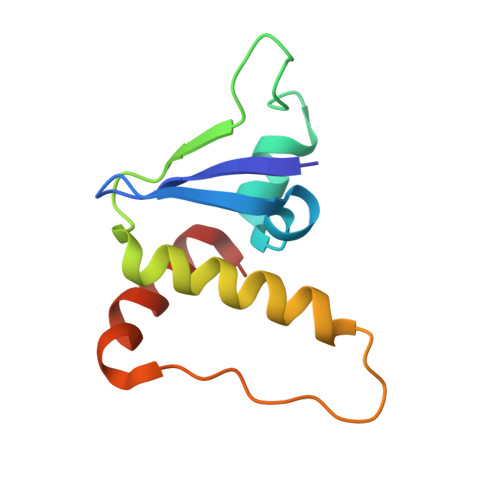
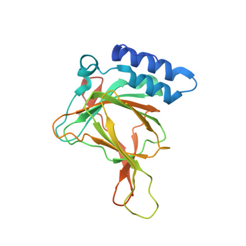
    The von Hippel-Lindau tumor suppressor gene product (pVHL) is an E3 ligase substrate receptor that binds proline-hydroxylated hypoxia-inducible factor HIF1α, leading to its ubiquitin-dependent degradation. By using protein arrays, we identified a small molecule that binds the HIF1α-binding pocket on pVHL and functions as a molecular glue degrader of the neosubstrate cysteine dioxygenase (CDO1) by recruiting it into the VHL-Cullin-RING E3 ligase complex and leading to its selective degradation. The CDO1-binding region involved in VHL recruitment was characterized through a combination of mutagenesis and protein-protein docking coupled with molecular-dynamics-based solvation analysis. The X-ray structure of the ternary complexes of VHL, CDO1 and degrader molecules confirms the binding region prediction and provides atomic insights into key molecular glue interactions.


  • Organizational Affiliation
    • Discovery Sciences, Novartis Biomedical Research, Cambridge, MA, USA.

Macromolecules
Find similar proteins by:  (by identity cutoff)  |  3D Structure
Entity ID: 1
MoleculeChains Sequence LengthOrganismDetailsImage
von Hippel-Lindau disease tumor suppressor162Homo sapiensMutation(s): 0 
Gene Names: VHL
UniProt & NIH Common Fund Data Resources
Find proteins for P40337 (Homo sapiens)
Explore P40337 
Go to UniProtKB:  P40337
PHAROS:  P40337
GTEx:  ENSG00000134086 
Entity Groups  
Sequence Clusters30% Identity50% Identity70% Identity90% Identity95% Identity100% Identity
UniProt GroupP40337
Sequence Annotations
Expand
  • Reference Sequence
Find similar proteins by:  (by identity cutoff)  |  3D Structure
Entity ID: 2
MoleculeChains Sequence LengthOrganismDetailsImage
Elongin-B118Homo sapiensMutation(s): 0 
Gene Names: ELOBTCEB2
UniProt & NIH Common Fund Data Resources
Find proteins for Q15370 (Homo sapiens)
Explore Q15370 
Go to UniProtKB:  Q15370
PHAROS:  Q15370
GTEx:  ENSG00000103363 
Entity Groups  
Sequence Clusters30% Identity50% Identity70% Identity90% Identity95% Identity100% Identity
UniProt GroupQ15370
Sequence Annotations
Expand
  • Reference Sequence
Find similar proteins by:  (by identity cutoff)  |  3D Structure
Entity ID: 3
MoleculeChains Sequence LengthOrganismDetailsImage
Elongin-C96Homo sapiensMutation(s): 0 
Gene Names: ELOCTCEB1
UniProt & NIH Common Fund Data Resources
Find proteins for Q15369 (Homo sapiens)
Explore Q15369 
Go to UniProtKB:  Q15369
PHAROS:  Q15369
GTEx:  ENSG00000154582 
Entity Groups  
Sequence Clusters30% Identity50% Identity70% Identity90% Identity95% Identity100% Identity
UniProt GroupQ15369
Sequence Annotations
Expand
  • Reference Sequence
Find similar proteins by:  (by identity cutoff)  |  3D Structure
Entity ID: 4
MoleculeChains Sequence LengthOrganismDetailsImage
Cysteine dioxygenase type 1201Homo sapiensMutation(s): 0 
Gene Names: CDO1
EC: 1.13.11.20
UniProt & NIH Common Fund Data Resources
Find proteins for Q16878 (Homo sapiens)
Explore Q16878 
Go to UniProtKB:  Q16878
PHAROS:  Q16878
GTEx:  ENSG00000129596 
Entity Groups  
Sequence Clusters30% Identity50% Identity70% Identity90% Identity95% Identity100% Identity
UniProt GroupQ16878
Sequence Annotations
Expand
  • Reference Sequence
Small Molecules
Binding Affinity Annotations 
IDSourceBinding Affinity
3JF BindingDB:  8VLB Kd: min: 107, max: 185 (nM) from 6 assay(s)
IC50: min: 185, max: 2540 (nM) from 6 assay(s)
Experimental Data & Validation

Experimental Data

  • Method: X-RAY DIFFRACTION
  • Resolution: 2.90 Å
  • R-Value Free:  0.258 (Depositor), 0.260 (DCC) 
  • R-Value Work:  0.208 (Depositor), 0.210 (DCC) 
  • R-Value Observed: 0.210 (Depositor) 
Space Group: C 2 2 21
Unit Cell:
Length ( Å )Angle ( ˚ )
a = 116.44α = 90
b = 178.116β = 90
c = 104.166γ = 90
Software Package:
Software NamePurpose
PHENIXrefinement
XDSdata reduction
XDSdata scaling
PHASERphasing

Structure Validation

View Full Validation Report


Ligand Structure Quality Assessment 


Entry History & Funding Information

Deposition Data


Funding OrganizationLocationGrant Number
Not funded--

Revision History  (Full details and data files)

  • Version 1.0: 2024-03-13
    Type: Initial release
  • Version 1.1: 2025-07-09
    Changes: Database references, Structure summary