8XIL | pdb_00008xil

Cellodextrin phosphorylase from Clostridium thermocellum mutant - all cysteine residues were substituted with serines


Experimental Data Snapshot

  • Method: X-RAY DIFFRACTION
  • Resolution: 1.21 Å
  • R-Value Free: 
    0.164 (Depositor), 0.164 (DCC) 
  • R-Value Work: 
    0.143 (Depositor), 0.143 (DCC) 
  • R-Value Observed: 
    0.144 (Depositor) 

Starting Model: in silico
View more details

wwPDB Validation   3D Report Full Report


Ligand Structure Quality Assessment 


This is version 1.1 of the entry. See complete history


Literature

Effect of Free Cysteine Residues to Serine Mutation on Cellodextrin Phosphorylase.

Kuga, T.Sunagawa, N.Igarashi, K.

(2024) J Appl Glycosci (1999) 71: 37-46

  • DOI: https://doi.org/10.5458/jag.jag.JAG-2023_0011
  • Primary Citation of Related Structures:  
    8XI1, 8XIL, 8XIS

  • PubMed Abstract: 

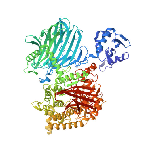
    Cellodextrin phosphorylase (CDP) plays a key role in energy-efficient cellulose metabolism of anaerobic bacteria by catalyzing phosphorolysis of cellodextrin to produce cellobiose and glucose 1-phosphate, which can be utilized for glycolysis without consumption of additional ATP. As the enzymatic phosphorolysis reaction is reversible, CDP is also employed to produce cellulosic materials in vitro. However, the enzyme is rapidly inactivated by oxidation, which hinders in vitro utilization in aerobic environments. It has been suggested that the cysteine residues of CDP, which do not form disulfide bonds, are responsible for the loss of activity, and the aim of the present work was to test this idea. For this purpose, we replaced all 11 free cysteine residues of CDP from Acetivibrio thermocellus (formerly known as Clostridium thermocellum ) with serine, which structurally resembles cysteine in our previous work. Herein, we show that the resulting CDP variant, named CDP-CS, has comparable activity to the wild-type enzyme, but shows increased stability to oxidation during long-term storage. X-Ray crystallography indicated that the mutations did not markedly alter the overall structure of the enzyme. Ensemble refinement of the crystal structures of CDP and CDP-CS indicated that the C372S and C625S mutations reduce structural fluctuations in the protein main chain, which may contribute to the increased stability of CDP-CS to oxidation.


  • Organizational Affiliation
    • 1 Department of Biomaterial Sciences, Graduate School of Agricultural and Life Sciences, The University of Tokyo.

Macromolecules
Find similar proteins by:  (by identity cutoff)  |  3D Structure
Entity ID: 1
MoleculeChains Sequence LengthOrganismDetailsImage
Cellodextrin phosphorylase
A, B
992Acetivibrio thermocellusMutation(s): 12 
Gene Names: cdp-ym4
UniProt
Find proteins for Q93HT8 (Acetivibrio thermocellus)
Explore Q93HT8 
Go to UniProtKB:  Q93HT8
Entity Groups  
Sequence Clusters30% Identity50% Identity70% Identity90% Identity95% Identity100% Identity
UniProt GroupQ93HT8
Sequence Annotations
Expand
  • Reference Sequence
Oligosaccharides

Help

Entity ID: 2
MoleculeChains Length2D Diagram Glycosylation3D Interactions
beta-D-glucopyranose-(1-4)-beta-D-glucopyranose-(1-4)-beta-D-glucopyranose
C, D
3N/A
Glycosylation Resources
GlyTouCan:  G14338VK
GlyCosmos:  G14338VK
GlyGen:  G14338VK
Small Molecules
Ligands 6 Unique
IDChains Name / Formula / InChI Key2D Diagram3D Interactions
TRS
Query on TRS

Download Ideal Coordinates CCD File 
BA [auth B],
EA [auth B],
G [auth A]
2-AMINO-2-HYDROXYMETHYL-PROPANE-1,3-DIOL
C4 H12 N O3
LENZDBCJOHFCAS-UHFFFAOYSA-O
PEG
Query on PEG

Download Ideal Coordinates CCD File 
HA [auth B],
IA [auth B],
L [auth A],
M [auth A],
N [auth A]
DI(HYDROXYETHYL)ETHER
C4 H10 O3
MTHSVFCYNBDYFN-UHFFFAOYSA-N
SO4 (Subject of Investigation/LOI)
Query on SO4

Download Ideal Coordinates CCD File 
GA [auth B],
K [auth A]
SULFATE ION
O4 S
QAOWNCQODCNURD-UHFFFAOYSA-L
GOL
Query on GOL

Download Ideal Coordinates CCD File 
JA [auth B]
KA [auth B]
LA [auth B]
MA [auth B]
NA [auth B]
JA [auth B],
KA [auth B],
LA [auth B],
MA [auth B],
NA [auth B],
O [auth A],
OA [auth B],
P [auth A],
PA [auth B],
Q [auth A],
QA [auth B],
R [auth A],
RA [auth B],
S [auth A],
SA [auth B],
T [auth A],
TA [auth B],
U [auth A],
UA [auth B],
V [auth A],
W [auth A],
X [auth A],
Y [auth A]
GLYCEROL
C3 H8 O3
PEDCQBHIVMGVHV-UHFFFAOYSA-N
ACT
Query on ACT

Download Ideal Coordinates CCD File 
CA [auth B]
DA [auth B]
E [auth A]
F [auth A]
FA [auth B]
CA [auth B],
DA [auth B],
E [auth A],
F [auth A],
FA [auth B],
H [auth A],
I [auth A],
J [auth A]
ACETATE ION
C2 H3 O2
QTBSBXVTEAMEQO-UHFFFAOYSA-M
CL
Query on CL

Download Ideal Coordinates CCD File 
AA [auth A],
VA [auth B],
WA [auth B],
XA [auth B],
Z [auth A]
CHLORIDE ION
Cl
VEXZGXHMUGYJMC-UHFFFAOYSA-M
Biologically Interesting Molecules (External Reference) 1 Unique
Experimental Data & Validation

Experimental Data

  • Method: X-RAY DIFFRACTION
  • Resolution: 1.21 Å
  • R-Value Free:  0.164 (Depositor), 0.164 (DCC) 
  • R-Value Work:  0.143 (Depositor), 0.143 (DCC) 
  • R-Value Observed: 0.144 (Depositor) 
Space Group: P 1
Unit Cell:
Length ( Å )Angle ( ˚ )
a = 83.208α = 98.576
b = 88.778β = 110.545
c = 88.761γ = 110.564
Software Package:
Software NamePurpose
PHENIXrefinement
XDSdata reduction
Aimlessdata scaling
PHENIXphasing

Structure Validation

View Full Validation Report


Ligand Structure Quality Assessment 


Entry History & Funding Information

Deposition Data


Funding OrganizationLocationGrant Number
Japan Society for the Promotion of Science (JSPS)Japan22J12566
Ministry of Education, Culture, Sports, Science and Technology (Japan)Japan18H05494

Revision History  (Full details and data files)

  • Version 1.0: 2024-12-25
    Type: Initial release
  • Version 1.1: 2025-07-09
    Changes: Database references