8YRJ | pdb_00008yrj

Mouse Fc epsilon RI


Experimental Data Snapshot

  • Method: ELECTRON MICROSCOPY
  • Resolution: 3.87 Å
  • Aggregation State: PARTICLE 
  • Reconstruction Method: SINGLE PARTICLE 

wwPDB Validation   3D Report Full Report


This is version 1.4 of the entry. See complete history


Literature

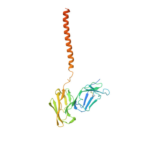
Architecture of the high-affinity immunoglobulin E receptor.

Zhang, Z.Yui, M.Ohto, U.Shimizu, T.

(2024) Sci Signal 17: eadn1303-eadn1303

  • DOI: https://doi.org/10.1126/scisignal.adn1303
  • Primary Citation of Related Structures:  
    8K7R, 8K7S, 8K7T, 8YRJ

  • PubMed Abstract: 

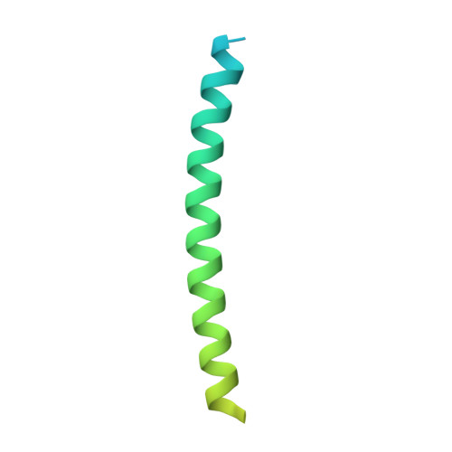
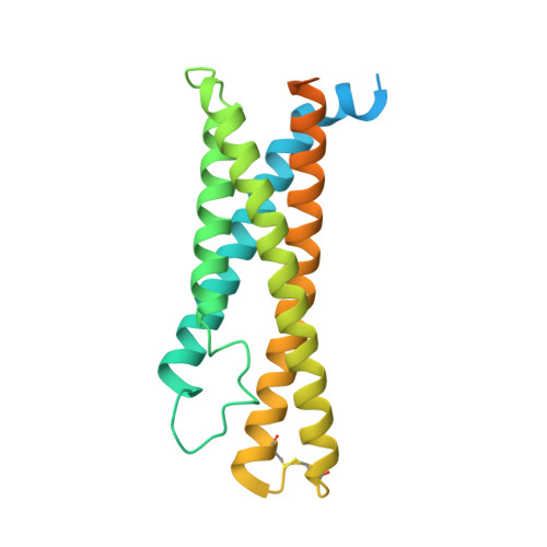
    The high-affinity immunoglobulin E (IgE) receptor (FcεRI) drives type I hypersensitivity in response to allergen-specific IgE. FcεRI is a multimeric complex typically composed of one α, one β, and two disulfide-linked γ subunits. The α subunit binds to the fragment crystallizable (Fc) region of IgE (Fcε), whereas the β and γ subunits mediate signaling through their intracellular immunoreceptor tyrosine-based activation motifs (ITAMs). Here, we report cryo-electron microscopy (cryo-EM) structures of the apo state of FcεRI and of FcεRI bound to Fcε. At the transmembrane domain (TMD), the α and γ subunits associate to form a tightly packed, three-helix bundle (αγ 2 bundle) with pseudo-threefold symmetry through extensive hydrophobic and polar interactions. The αγ 2 bundle further assembles with the β subunit to complete the TMD, from which multiple ITAMs might extend into the cytoplasm for downstream signaling. The apo mouse FcεRI essentially forms an identical structure to that of the Fcε-bound sensitized form, suggesting that the binding of Fcε to FcεRI does not alter the overall conformation of the receptor. Furthermore, the juxtamembrane interaction between the extracellular domains (ECDs) of mouse FcεRIα and FcεRIβ is not observed between their human counterparts, which implies potential species-specific differences in receptor stability and activation. Our findings provide a framework for understanding the general structural principles underlying Fc receptor assembly, the signaling mechanism underlying type I hypersensitivity, and the design of efficient antiallergic therapeutics.


  • Organizational Affiliation
    • Graduate School of Pharmaceutical Sciences, University of Tokyo, 7-3-1 Hongo, Bunkyo-ku, Tokyo 113-0033, Japan.

Macromolecules
Find similar proteins by:  (by identity cutoff)  |  3D Structure
Entity ID: 1
MoleculeChains Sequence LengthOrganismDetailsImage
High affinity immunoglobulin epsilon receptor subunit alpha273Mus musculusMutation(s): 0 
Gene Names: Fcer1aFce1a
Membrane Entity: Yes 
UniProt
Find proteins for P20489 (Mus musculus)
Explore P20489 
Go to UniProtKB:  P20489
Entity Groups  
Sequence Clusters30% Identity50% Identity70% Identity90% Identity95% Identity100% Identity
UniProt GroupP20489
Glycosylation
Glycosylation Sites: 5Go to GlyGen: P20489-1
Sequence Annotations
Expand
  • Reference Sequence
Find similar proteins by:  (by identity cutoff)  |  3D Structure
Entity ID: 2
MoleculeChains Sequence LengthOrganismDetailsImage
High affinity immunoglobulin epsilon receptor subunit gammaB [auth G],
C [auth H]
104Mus musculusMutation(s): 0 
Gene Names: Fcer1gFce1g
Membrane Entity: Yes 
UniProt & NIH Common Fund Data Resources
Find proteins for P20491 (Mus musculus)
Explore P20491 
Go to UniProtKB:  P20491
IMPC:  MGI:95496
Entity Groups  
Sequence Clusters30% Identity50% Identity70% Identity90% Identity95% Identity100% Identity
UniProt GroupP20491
Sequence Annotations
Expand
  • Reference Sequence
Find similar proteins by:  (by identity cutoff)  |  3D Structure
Entity ID: 3
MoleculeChains Sequence LengthOrganismDetailsImage
High affinity immunoglobulin epsilon receptor subunit betaD [auth B]245Mus musculusMutation(s): 0 
Gene Names: Ms4a2Fce1bFcer1bMs4a1
Membrane Entity: Yes 
UniProt
Find proteins for P20490 (Mus musculus)
Explore P20490 
Go to UniProtKB:  P20490
Entity Groups  
Sequence Clusters30% Identity50% Identity70% Identity90% Identity95% Identity100% Identity
UniProt GroupP20490
Sequence Annotations
Expand
  • Reference Sequence
Oligosaccharides

Help

Entity ID: 4
MoleculeChains Length2D Diagram Glycosylation3D Interactions
beta-D-mannopyranose-(1-4)-2-acetamido-2-deoxy-beta-D-glucopyranose-(1-4)-2-acetamido-2-deoxy-beta-D-glucopyranoseE [auth C],
F [auth D]
3N-Glycosylation
Glycosylation Resources
GlyTouCan:  G15407YE
GlyCosmos:  G15407YE
GlyGen:  G15407YE
Experimental Data & Validation

Experimental Data

  • Method: ELECTRON MICROSCOPY
  • Resolution: 3.87 Å
  • Aggregation State: PARTICLE 
  • Reconstruction Method: SINGLE PARTICLE 
EM Software:
TaskSoftware PackageVersion
MODEL REFINEMENTPHENIX

Structure Validation

View Full Validation Report


Entry History & Funding Information

Deposition Data


Funding OrganizationLocationGrant Number
Not funded--

Revision History  (Full details and data files)

  • Version 1.0: 2024-07-17
    Type: Initial release
  • Version 1.1: 2024-11-13
    Changes: Data collection, Structure summary
  • Version 1.2: 2024-11-27
    Changes: Data collection
  • Version 1.3: 2025-01-29
    Changes: Data collection, Database references
  • Version 1.4: 2025-06-25
    Changes: Data collection