8ZZL | pdb_00008zzl

Structure of complement C3(H2O) revealed by quantitative cross-linking/mass spectrometry and modeling

Integrative structure models are generated using different types of input information, including varied experimental data, physical principles, statistical preferences, and other prior information.


Integrative Structure Snapshot

  • Multi-Scale: Yes 
  • Multi-State: Yes 
  • Ordered-State: Yes 
  • Deposited Models: 4 
  • Representative Model: 1 

This is version 1.0 of the entry. See complete history


Literature

Structure of Complement C3(H2O) Revealed By Quantitative Cross-Linking/Mass Spectrometry And Modeling.

Chen, Z.A.Pellarin, R.Fischer, L.Sali, A.Nilges, M.Barlow, P.N.Rappsilber, J.

(2016) Mol Cell Proteomics 15: 2730-2743

  • DOI: https://doi.org/10.1074/mcp.M115.056473
  • Primary Citation of Related Structures:  
    8ZZL

  • PubMed Abstract: 

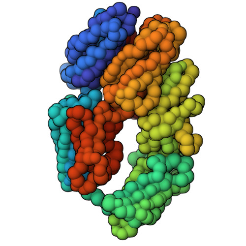
    The slow but spontaneous and ubiquitous formation of C3(H2O), the hydrolytic and conformationally rearranged product of C3, initiates antibody-independent activation of the complement system that is a key first line of antimicrobial defense. The structure of C3(H2O) has not been determined. Here we subjected C3(H2O) to quantitative cross-linking/mass spectrometry (QCLMS). This revealed details of the structural differences and similarities between C3(H2O) and C3, as well as between C3(H2O) and its pivotal proteolytic cleavage product, C3b, which shares functionally similarity with C3(H2O). Considered in combination with the crystal structures of C3 and C3b, the QCMLS data suggest that C3(H2O) generation is accompanied by the migration of the thioester-containing domain of C3 from one end of the molecule to the other. This creates a stable C3b-like platform able to bind the zymogen, factor B, or the regulator, factor H. Integration of available crystallographic and QCLMS data allowed the determination of a 3D model of the C3(H2O) domain architecture. The unique arrangement of domains thus observed in C3(H2O), which retains the anaphylatoxin domain (that is excised when C3 is enzymatically activated to C3b), can be used to rationalize observed differences between C3(H2O) and C3b in terms of complement activation and regulation.


  • Organizational Affiliation
    • From the ‡Wellcome Trust Centre for Cell Biology, Institute of Cell Biology, School of Biological Sciences, University of Edinburgh, Edinburgh EH9 3BF, UK;

Macromolecules
Find similar proteins by:  (by identity cutoff) 
Entity ID: 1
MoleculeChains Sequence LengthOrganismDetailsImage
beta645Homo sapiensMutation(s): 0 
Gene Names: C3
UniProt & NIH Common Fund Data Resources
Find proteins for P01024 (Homo sapiens)
Explore P01024 
Go to UniProtKB:  P01024
PHAROS:  P01024
GTEx:  ENSG00000125730 
Entity Groups  
Sequence Clusters30% Identity50% Identity70% Identity90% Identity95% Identity100% Identity
UniProt GroupP01024
Sequence Annotations
Expand
  • Reference Sequence
Find similar proteins by:  (by identity cutoff) 
Entity ID: 2
MoleculeChains Sequence LengthOrganismDetailsImage
alpha992Homo sapiensMutation(s): 0 
Gene Names: C3
UniProt & NIH Common Fund Data Resources
Find proteins for P01024 (Homo sapiens)
Explore P01024 
Go to UniProtKB:  P01024
PHAROS:  P01024
GTEx:  ENSG00000125730 
Entity Groups  
Sequence Clusters30% Identity50% Identity70% Identity90% Identity95% Identity100% Identity
UniProt GroupP01024
Sequence Annotations
Expand
  • Reference Sequence
Experimental Data & Validation

Integrative Structure Snapshot

  • Multi-Scale: Yes 
  • Multi-State: Yes 
  • Ordered-State: Yes 
  • Deposited Models: 4 
  • Representative Model: 1 

Structure Validation

View Full Validation Report

View Summary Validation Report


Entry History 

Deposition Data

Revision History  (Full details and data files)

  • Version 1.0: 2018-06-14
    Type: Initial release