8F8O | pdb_00008f8o

Crystal Structure of the Succinyl-diaminopimelate Desuccinylase (DapE) from Acinetobacter baumannii in complex with Succinic and L-Lactic Acids


Experimental Data Snapshot

  • Method: X-RAY DIFFRACTION
  • Resolution: 2.10 Å
  • R-Value Free: 
    0.230 (Depositor), 0.240 (DCC) 
  • R-Value Work: 
    0.189 (Depositor), 0.200 (DCC) 
  • R-Value Observed: 
    0.191 (Depositor) 

Starting Model: experimental
View more details

wwPDB Validation   3D Report Full Report


Ligand Structure Quality Assessment 


This is version 1.3 of the entry. See complete history


Literature

Biochemical and Structural Analysis of the Bacterial Enzyme Succinyl-Diaminopimelate Desuccinylase (DapE) from Acinetobacter baumannii.

Kelley, E.H.Minasov, G.Konczak, K.Shuvalova, L.Brunzelle, J.S.Shukla, S.Beulke, M.Thabthimthong, T.Olsen, K.W.Inniss, N.L.Satchell, K.J.F.Becker, D.P.

(2024) ACS Omega 9: 3905-3915

  • DOI: https://doi.org/10.1021/acsomega.3c08231
  • Primary Citation of Related Structures:  
    8F8O

  • PubMed Abstract: 

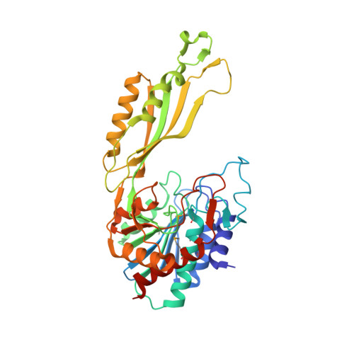
    There is an urgent need for new antibiotics given the rise of antibiotic resistance, and succinyl-diaminopimelate desuccinylase (DapE, E.C. 3.5.1.18) has emerged as a promising bacterial enzyme target. DapE from Haemophilus influenzae ( Hi DapE) has been studied and inhibitors identified, but it is essential to explore DapE from different species to assess selective versus broad-spectrum therapeutics. We have determined the structure of DapE from the ESKAPE pathogen Acinetobacter baumannii ( Ab DapE) and studied inhibition by known inhibitors of Hi DapE. Ab DapE is inhibited by captopril and sulfate comparable to Hi DapE, but Ab DapE was not significantly inhibited by a known indoline sulfonamide Hi DapE inhibitor. Captopril and sulfate both stabilize Hi DapE by increasing the thermal melting temperature ( T m ) in thermal shift assays. By contrast, sulfate decreases the stability of the Ab DapE enzyme, whereas captopril increases the stability. Further, we report two crystal structures of selenomethionine-substituted Ab DapE in the closed conformation, one with Ab DapE in complex with succinate derived from enzymatic hydrolysis of N 6 -methyl-l,l-SDAP substrate and acetate (PDB code 7T1Q, 2.25 Å resolution), and a crystal structure of Ab DapE with bound succinate along with l-(S)-lactate, a product of degradation of citric acid from the crystallization buffer during X-ray irradiation (PDB code 8F8O, 2.10 Å resolution).


  • Organizational Affiliation
    • Department of Chemistry and Biochemistry, Loyola University Chicago, 1032 West Sheridan Road, Chicago, Illinois 60660, United States.

Macromolecules
Find similar proteins by:  (by identity cutoff)  |  3D Structure
Entity ID: 1
MoleculeChains Sequence LengthOrganismDetailsImage
Succinyl-diaminopimelate desuccinylase
A, B
384Acinetobacter baumannii ATCC 17978Mutation(s): 0 
Gene Names: dapEA1S_2810
EC: 3.5.1.18
UniProt
Find proteins for A3M8H2 (Acinetobacter baumannii (strain ATCC 17978 / DSM 105126 / CIP 53.77 / LMG 1025 / NCDC KC755 / 5377))
Explore A3M8H2 
Go to UniProtKB:  A3M8H2
Entity Groups  
Sequence Clusters30% Identity50% Identity70% Identity90% Identity95% Identity100% Identity
UniProt GroupA3M8H2
Sequence Annotations
Expand
  • Reference Sequence
Small Molecules
Ligands 6 Unique
IDChains Name / Formula / InChI Key2D Diagram3D Interactions
PG4
Query on PG4

Download Ideal Coordinates CCD File 
L [auth B]TETRAETHYLENE GLYCOL
C8 H18 O5
UWHCKJMYHZGTIT-UHFFFAOYSA-N
CIT
Query on CIT

Download Ideal Coordinates CCD File 
M [auth B]CITRIC ACID
C6 H8 O7
KRKNYBCHXYNGOX-UHFFFAOYSA-N
PGE
Query on PGE

Download Ideal Coordinates CCD File 
G [auth A]TRIETHYLENE GLYCOL
C6 H14 O4
ZIBGPFATKBEMQZ-UHFFFAOYSA-N
SIN (Subject of Investigation/LOI)
Query on SIN

Download Ideal Coordinates CCD File 
E [auth A],
J [auth B]
SUCCINIC ACID
C4 H6 O4
KDYFGRWQOYBRFD-UHFFFAOYSA-N
2OP (Subject of Investigation/LOI)
Query on 2OP

Download Ideal Coordinates CCD File 
F [auth A],
K [auth B]
(2S)-2-HYDROXYPROPANOIC ACID
C3 H6 O3
JVTAAEKCZFNVCJ-REOHCLBHSA-N
ZN (Subject of Investigation/LOI)
Query on ZN

Download Ideal Coordinates CCD File 
C [auth A],
D [auth A],
H [auth B],
I [auth B]
ZINC ION
Zn
PTFCDOFLOPIGGS-UHFFFAOYSA-N
Modified Residues  1 Unique
IDChains TypeFormula2D DiagramParent
MSE
Query on MSE
A, B
L-PEPTIDE LINKINGC5 H11 N O2 SeMET
Experimental Data & Validation

Experimental Data

  • Method: X-RAY DIFFRACTION
  • Resolution: 2.10 Å
  • R-Value Free:  0.230 (Depositor), 0.240 (DCC) 
  • R-Value Work:  0.189 (Depositor), 0.200 (DCC) 
  • R-Value Observed: 0.191 (Depositor) 
Space Group: P 21 21 21
Unit Cell:
Length ( Å )Angle ( ˚ )
a = 67.659α = 90
b = 102.533β = 90
c = 145.491γ = 90
Software Package:
Software NamePurpose
REFMACrefinement
HKL-3000data reduction
HKL-3000data scaling
PHASERphasing

Structure Validation

View Full Validation Report


Ligand Structure Quality Assessment 


Entry History & Funding Information

Deposition Data


Funding OrganizationLocationGrant Number
National Institutes of Health/National Institute Of Allergy and Infectious Diseases (NIH/NIAID)United States75N93022C00035

Revision History  (Full details and data files)

  • Version 1.0: 2022-11-30
    Type: Initial release
  • Version 1.1: 2023-02-01
    Changes: Database references
  • Version 1.2: 2023-02-22
    Changes: Data collection, Experimental preparation
  • Version 1.3: 2025-09-24
    Changes: Data collection, Database references, Structure summary