9B3M | pdb_00009b3m

NorA in inward-open conformation (NorA-BRIL fusion)

  • Classification: TRANSPORT PROTEIN
  • Organism(s): Staphylococcus aureus
  • Expression System: Escherichia coli BL21(DE3)
  • Mutation(s): No 
  • Membrane Protein: Yes  PDBTMMemProtMDmpstruc

  • Deposited: 2024-03-19 Released: 2024-12-11 
  • Deposition Author(s): Xie, P., Li, Y., Kuang, H., Wang, D.N., Traaseth, N.J.
  • Funding Organization(s): National Institutes of Health/National Institute Of Allergy and Infectious Diseases (NIH/NIAID), National Science Foundation (NSF, United States), National Institutes of Health/National Institute of Neurological Disorders and Stroke (NIH/NINDS), National Institutes of Health/National Institute of Diabetes and Digestive and Kidney Disease (NIH/NIDDK)

Experimental Data Snapshot

  • Method: ELECTRON MICROSCOPY
  • Resolution: 3.21 Å
  • Aggregation State: PARTICLE 
  • Reconstruction Method: SINGLE PARTICLE 

Starting Model: other
View more details

wwPDB Validation   3D Report Full Report


This is version 1.1 of the entry. See complete history


Literature

A fiducial-assisted strategy compatible with resolving small MFS transporter structures in multiple conformations using cryo-EM.

Xie, P.Li, Y.Lamon, G.Kuang, H.Wang, D.N.Traaseth, N.J.

(2025) Nat Commun 16: 7-7

  • DOI: https://doi.org/10.1038/s41467-024-54986-5
  • Primary Citation of Related Structures:  
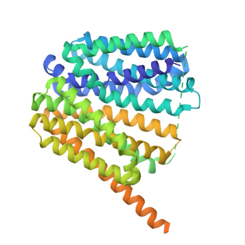
    9B3K, 9B3L, 9B3M, 9B3O

  • PubMed Abstract: 

    Advancements in cryo-EM have stimulated a revolution in structural biology. Yet, for membrane proteins near the cryo-EM size threshold of approximately 40 kDa, including transporters and G-protein coupled receptors, the absence of distinguishable structural features makes image alignment and structure determination a significant challenge. Furthermore, resolving more than one protein conformation within a sample, a major advantage of cryo-EM, represents an even greater degree of difficulty. Here, we describe a strategy for introducing a rigid fiducial marker (BRIL domain) at the C-terminus of membrane transporters from the Major Facilitator Superfamily (MFS) with AlphaFold2. This approach involves fusion of the last transmembrane domain helix of the target protein with the first helix of BRIL through a short poly-alanine linker to promote helicity. Combining this strategy with a BRIL-specific Fab, we elucidated four cryo-EM structures of the 42 kDa Staphylococcus aureus transporter NorA, three of which were derived from a single sample corresponding to inward-open, inward-occluded, and occluded conformations. Hence, this fusion construct facilitated experiments to characterize the conformational landscape of NorA and validated our design to position the BRIL/antibody pair in an orientation that avoids steric clash with the transporter. The latter was enabled through AlphaFold2 predictions, which minimized guesswork and reduced the need for screening several constructs. We further validated the suitability of the method to three additional MFS transporters (GlpT, Bmr, and Blt), results that supported a rigid linker between the transporter and BRIL. The successful application to four MFS proteins, the largest family of secondary transporters in nature, and analysis of predicted structures for the family indicates this strategy will be a valuable tool for studying other MFS members using cryo-EM.


  • Organizational Affiliation
    • Department of Chemistry, New York University, New York, NY, USA.

Macromolecules
Find similar proteins by:  (by identity cutoff)  |  3D Structure
Entity ID: 1
MoleculeChains Sequence LengthOrganismDetailsImage
NorA-BRIL(3A) fusion489Staphylococcus aureusMutation(s): 0 
Gene Names: norA
Membrane Entity: Yes 
UniProt
Find proteins for Q2G0A2 (Staphylococcus aureus (strain NCTC 8325 / PS 47))
Explore Q2G0A2 
Go to UniProtKB:  Q2G0A2
Entity Groups  
Sequence Clusters30% Identity50% Identity70% Identity90% Identity95% Identity100% Identity
UniProt GroupQ2G0A2
Sequence Annotations
Expand
  • Reference Sequence
Experimental Data & Validation

Experimental Data

  • Method: ELECTRON MICROSCOPY
  • Resolution: 3.21 Å
  • Aggregation State: PARTICLE 
  • Reconstruction Method: SINGLE PARTICLE 
EM Software:
TaskSoftware PackageVersion
MODEL REFINEMENTPHENIX1.20.1
RECONSTRUCTIONcryoSPARCv3.3.1

Structure Validation

View Full Validation Report


Entry History & Funding Information

Deposition Data


Funding OrganizationLocationGrant Number
National Institutes of Health/National Institute Of Allergy and Infectious Diseases (NIH/NIAID)United StatesR01AI165782
National Institutes of Health/National Institute Of Allergy and Infectious Diseases (NIH/NIAID)United StatesAI108889
National Science Foundation (NSF, United States)United StatesMCB1902449
National Institutes of Health/National Institute of Neurological Disorders and Stroke (NIH/NINDS)United StatesR01NS108151
National Institutes of Health/National Institute of Diabetes and Digestive and Kidney Disease (NIH/NIDDK)United StatesR01DK135088

Revision History  (Full details and data files)

  • Version 1.0: 2024-12-11
    Type: Initial release
  • Version 1.1: 2025-06-25
    Changes: Data collection, Database references