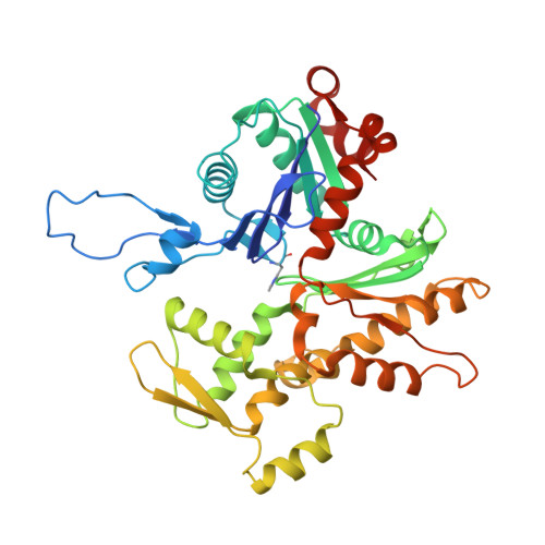
The hypertrophic cardiomyopathy-associated A331P actin variant enhances basal contractile activity and elicits resting muscle dysfunction.
Doran, M.H., Rynkiewicz, M.J., Despond, E., Viswanathan, M.C., Madan, A., Chitre, K., Fenwick, A.J., Sousa, D., Lehman, W., Dawson, J.F., Cammarato, A.(2025) iScience 28: 111816-111816
- PubMed: 39981516
- DOI: https://doi.org/10.1016/j.isci.2025.111816
- Primary Citation of Related Structures:
9B3R - PubMed Abstract:
Previous studies aimed at defining the mechanistic basis of hypertrophic cardiomyopathy caused by A331P cardiac actin have reported conflicting results. The mutation is located along an actin surface strand, proximal to residues that interact with tropomyosin. These F-actin-tropomyosin associations are vital for proper contractile inhibition. To help resolve disease pathogenesis, we implemented a multidisciplinary approach. Transgenic Drosophila , expressing A331P actin, displayed skeletal muscle hypercontraction and elevated basal myocardial activity. A331P thin filaments, reconstituted using recombinant human cardiac actin, exhibited higher in vitro myosin-based sliding speeds, exclusively at low Ca 2+ concentrations. Cryo-EM-based reconstructions revealed no detectable A331P-related structural perturbations in F-actin. In silico , however, the P331-containing actin surface strand was less mobile and established diminished van der Waal's attractive forces with tropomyosin, which correlated with greater variability in inhibitory tropomyosin positioning. Such mutation-induced effects potentially elevate resting contractile activity among our models and may stimulate pathology in patients.
- Department of Pharmacology, Physiology & Biophysics, Boston University Chobanian & Avedisian School of Medicine, 72 E. Concord St, Boston, MA 02118, USA.
Organizational Affiliation:


