9BBO | pdb_00009bbo

Proline utilization A complexed with the product L-glutamate in the aldehyde dehydrogenase active site


Experimental Data Snapshot

  • Method: X-RAY DIFFRACTION
  • Resolution: 1.50 Å
  • R-Value Free: 
    0.191 (Depositor), 0.191 (DCC) 
  • R-Value Work: 
    0.169 (Depositor), 0.169 (DCC) 
  • R-Value Observed: 
    0.171 (Depositor) 

Starting Model: experimental
View more details

wwPDB Validation   3D Report Full Report


Ligand Structure Quality Assessment 


This is version 1.1 of the entry. See complete history


Literature

Visualization of Covalent Intermediates and Conformational States of Proline Utilization A by X-ray Crystallography and Molecular Dynamics Simulations.

Buckley, D.P.Becker, D.F.Tanner, J.J.

(2025) J Biological Chem : 110532-110532

  • DOI: https://doi.org/10.1016/j.jbc.2025.110532
  • Primary Citation of Related Structures:  
    9BBO, 9C34, 9C35, 9C36

  • PubMed Abstract: 

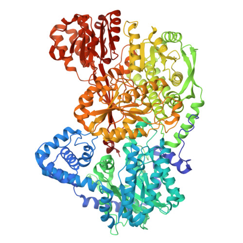
    The bifunctional enzyme proline utilization A (PutA) catalyzes the two-step oxidation of L-proline to L-glutamate using proline dehydrogenase (PRODH) and L-glutamate-γ-semialdehyde dehydrogenase (GSALDH) domains. The two active sites are 42 Å apart and connected by a buried tunnel that is hypothesized to channel the intermediates Δ 1 -pyrroline-5-carboxylate (P5C) and/or L-glutamate-γ-semialdehyde (GSAL). Kinetic and conventional X-ray crystallography of PutA from Sinorhizobium meliloti (SmPutA) were used to capture high resolution (1.47 - 1.88 Å) structures of states along the catalytic cycle, including a novel FADH - -proline covalent adduct in the PRODH site, the intermediate P5C bound noncovalently in the reduced PRODH active site, the covalent acyl-enzyme intermediate of the GSALDH reaction, and noncovalent complexes of GSAL and the final product L-glutamate in the GSALDH active site. The FADH - -proline covalent adduct resembles a stable species predicted from quantum mechanical electronic structure calculations of the PRODH reaction. The GSALDH domain complexes are consistent with conservation of substrate recognition and catalytic mechanism by the aldehyde dehydrogenase superfamily. The structure of reduced SmPutA with the P5C bound in the PRODH active site was used as the starting point for molecular dynamics simulations (21 x 2μs trajectories). P5C diffuses from the PRODH active site into the tunnel in most of the trajectories, but rarely dissociates completely from the enzyme, consistent with previous kinetic evidence of a substrate channeling mechanism. The simulations also provide insight into protein conformational changes associated with substrate channeling, including the opening and closing of a conserved ion pair gate.


  • Organizational Affiliation

    Department of Biochemistry, University of Missouri, Columbia, Missouri 65211, United States.


Macromolecules
Find similar proteins by:  (by identity cutoff)  |  3D Structure
Entity ID: 1
MoleculeChains Sequence LengthOrganismDetailsImage
Trifunctional transcriptional regulator/proline dehydrogenase/L-glutamate gamma-semialdehyde dehydrogenase
A, B
1,235Sinorhizobium melilotiMutation(s): 0 
Gene Names: putAGHK55_27665
EC: 1.2.1.88 (UniProt), 1.5.5.2 (UniProt)
UniProt
Find proteins for P95629 (Rhizobium meliloti)
Explore P95629 
Go to UniProtKB:  P95629
Entity Groups  
Sequence Clusters30% Identity50% Identity70% Identity90% Identity95% Identity100% Identity
UniProt GroupP95629
Sequence Annotations
Expand
  • Reference Sequence
Small Molecules
Ligands 5 Unique
IDChains Name / Formula / InChI Key2D Diagram3D Interactions
FAD (Subject of Investigation/LOI)
Query on FAD

Download Ideal Coordinates CCD File 
J [auth A],
R [auth B]
FLAVIN-ADENINE DINUCLEOTIDE
C27 H33 N9 O15 P2
VWWQXMAJTJZDQX-UYBVJOGSSA-N
PGE
Query on PGE

Download Ideal Coordinates CCD File 
I [auth A],
Q [auth B]
TRIETHYLENE GLYCOL
C6 H14 O4
ZIBGPFATKBEMQZ-UHFFFAOYSA-N
GGL (Subject of Investigation/LOI)
Query on GGL

Download Ideal Coordinates CCD File 
C [auth A],
K [auth B]
GAMMA-L-GLUTAMIC ACID
C5 H9 N O4
WHUUTDBJXJRKMK-VKHMYHEASA-N
PEG
Query on PEG

Download Ideal Coordinates CCD File 
P [auth B]DI(HYDROXYETHYL)ETHER
C4 H10 O3
MTHSVFCYNBDYFN-UHFFFAOYSA-N
SO4
Query on SO4

Download Ideal Coordinates CCD File 
D [auth A]
E [auth A]
F [auth A]
G [auth A]
H [auth A]
D [auth A],
E [auth A],
F [auth A],
G [auth A],
H [auth A],
L [auth B],
M [auth B],
N [auth B],
O [auth B]
SULFATE ION
O4 S
QAOWNCQODCNURD-UHFFFAOYSA-L
Experimental Data & Validation

Experimental Data

  • Method: X-RAY DIFFRACTION
  • Resolution: 1.50 Å
  • R-Value Free:  0.191 (Depositor), 0.191 (DCC) 
  • R-Value Work:  0.169 (Depositor), 0.169 (DCC) 
  • R-Value Observed: 0.171 (Depositor) 
Space Group: P 1 21 1
Unit Cell:
Length ( Å )Angle ( ˚ )
a = 101.377α = 90
b = 102.435β = 106.38
c = 127.598γ = 90
Software Package:
Software NamePurpose
PHENIXrefinement
Aimlessdata scaling
XDSdata reduction
PHENIXphasing

Structure Validation

View Full Validation Report


Ligand Structure Quality Assessment 


Entry History & Funding Information

Deposition Data

  • Released Date: 2025-03-12 
  • Deposition Author(s): Tanner, J.J.

Funding OrganizationLocationGrant Number
National Institutes of Health/National Institute of General Medical Sciences (NIH/NIGMS)United StatesR01GM065546

Revision History  (Full details and data files)

  • Version 1.0: 2025-03-12
    Type: Initial release
  • Version 1.1: 2025-08-13
    Changes: Database references