9E0Z | pdb_00009e0z

Dimeric motor domains from phi-like dynein-1 bound to a Lis1 dimer under Nde1-Lis1 condition

  • Classification: MOTOR PROTEIN
  • Organism(s): Homo sapiens
  • Expression System: Spodoptera frugiperda
  • Mutation(s): No 

  • Deposited: 2024-10-21 Released: 2025-08-06 
  • Deposition Author(s): Yang, J., Zhang, K.
  • Funding Organization(s): National Institutes of Health/National Institute of General Medical Sciences (NIH/NIGMS)

Experimental Data Snapshot

  • Method: ELECTRON MICROSCOPY
  • Resolution: 2.86 Å
  • Aggregation State: PARTICLE 
  • Reconstruction Method: SINGLE PARTICLE 

wwPDB Validation   3D Report Full Report


This is version 1.1 of the entry. See complete history


Literature

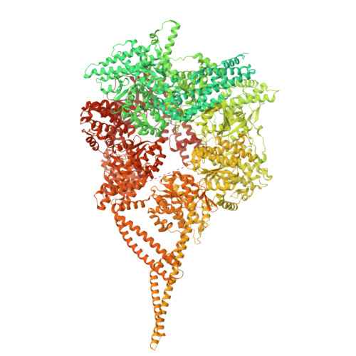
Nde1 promotes Lis1 binding to full-length autoinhibited human dynein 1.

Yang, J.Zhao, Y.Chai, P.Yildiz, A.Zhang, K.

(2025) Nat Chem Biol 

  • DOI: https://doi.org/10.1038/s41589-025-01981-6
  • Primary Citation of Related Structures:  
    9E0Z, 9E10, 9E11, 9E12, 9E13, 9E14

  • PubMed Abstract: 

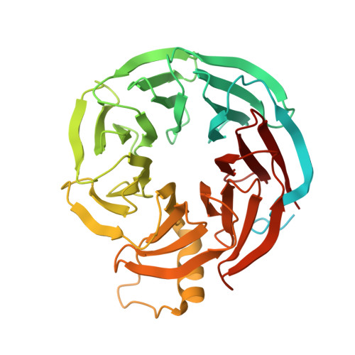
    Cytoplasmic dynein 1 (dynein) is the primary motor responsible for the retrograde transport of intracellular cargoes along microtubules. Activation of dynein requires the opening its autoinhibited Phi conformation, a process driven by Lis1 and Nde1/Ndel1. Using biochemical reconstitution and cryo-electron microscopy, we demonstrate that Nde1 enhances Lis1 binding to autoinhibited dynein and facilitates Phi opening. We identify a key intermediate in this activation pathway where a single Lis1 dimer binds between Phi-like (Phi L ) motor rings. In this 'Phi L -Lis1' complex, Lis1 interacts with one motor domain through canonical sites at the AAA+ (adenosine triphosphatases associated with diverse cellular activities) ring and stalk, and with AAA5, AAA6 and linker regions of the other motor domain. Mutagenesis and motility assays confirm the critical role of the Phi L -Lis1 interface in dynein activation. This intermediate forms rapidly in the presence of Nde1, although Nde1 is not part of Phi L -Lis1. These findings provide key insights into how Nde1 promotes Lis1-mediated Phi opening.


  • Organizational Affiliation
    • Department of Molecular Biophysics and Biochemistry, Yale University, New Haven, CT, USA.

Macromolecules
Find similar proteins by:  (by identity cutoff)  |  3D Structure
Entity ID: 1
MoleculeChains Sequence LengthOrganismDetailsImage
Cytoplasmic dynein 1 heavy chain 1
A, B
4,646Homo sapiensMutation(s): 0 
Gene Names: DYNC1H1DHC1DNCH1DNCLDNECLDYHCKIAA0325
UniProt & NIH Common Fund Data Resources
Find proteins for Q14204 (Homo sapiens)
Explore Q14204 
Go to UniProtKB:  Q14204
PHAROS:  Q14204
GTEx:  ENSG00000197102 
Entity Groups  
Sequence Clusters30% Identity50% Identity70% Identity90% Identity95% Identity100% Identity
UniProt GroupQ14204
Sequence Annotations
Expand
  • Reference Sequence
Find similar proteins by:  (by identity cutoff)  |  3D Structure
Entity ID: 2
MoleculeChains Sequence LengthOrganismDetailsImage
Platelet-activating factor acetylhydrolase IB subunit beta
C, D
410Homo sapiensMutation(s): 0 
Gene Names: PAFAH1B1LIS1MDCRMDSPAFAHA
UniProt & NIH Common Fund Data Resources
Find proteins for P43034 (Homo sapiens)
Explore P43034 
Go to UniProtKB:  P43034
PHAROS:  P43034
GTEx:  ENSG00000007168 
Entity Groups  
Sequence Clusters30% Identity50% Identity70% Identity90% Identity95% Identity100% Identity
UniProt GroupP43034
Sequence Annotations
Expand
  • Reference Sequence
Small Molecules
Ligands 3 Unique
IDChains Name / Formula / InChI Key2D Diagram3D Interactions
ATP (Subject of Investigation/LOI)
Query on ATP

Download Ideal Coordinates CCD File 
F [auth A],
L [auth B]
ADENOSINE-5'-TRIPHOSPHATE
C10 H16 N5 O13 P3
ZKHQWZAMYRWXGA-KQYNXXCUSA-N
ADP (Subject of Investigation/LOI)
Query on ADP

Download Ideal Coordinates CCD File 
E [auth A]
G [auth A]
H [auth A]
K [auth B]
M [auth B]
E [auth A],
G [auth A],
H [auth A],
K [auth B],
M [auth B],
N [auth B]
ADENOSINE-5'-DIPHOSPHATE
C10 H15 N5 O10 P2
XTWYTFMLZFPYCI-KQYNXXCUSA-N
MG (Subject of Investigation/LOI)
Query on MG

Download Ideal Coordinates CCD File 
I [auth A],
J [auth A],
O [auth B],
P [auth B]
MAGNESIUM ION
Mg
JLVVSXFLKOJNIY-UHFFFAOYSA-N
Experimental Data & Validation

Experimental Data

  • Method: ELECTRON MICROSCOPY
  • Resolution: 2.86 Å
  • Aggregation State: PARTICLE 
  • Reconstruction Method: SINGLE PARTICLE 
EM Software:
TaskSoftware PackageVersion
MODEL REFINEMENTPHENIX

Structure Validation

View Full Validation Report


Entry History & Funding Information

Deposition Data


Funding OrganizationLocationGrant Number
National Institutes of Health/National Institute of General Medical Sciences (NIH/NIGMS)United StatesR35 GM142959

Revision History  (Full details and data files)

  • Version 1.0: 2025-08-06
    Type: Initial release
  • Version 1.1: 2025-08-13
    Changes: Data collection, Database references