9FYA | pdb_00009fya

Structure of the Sabia Virus spike complex in a closed conformation


Experimental Data Snapshot

  • Method: ELECTRON MICROSCOPY
  • Resolution: 2.64 Å
  • Aggregation State: PARTICLE 
  • Reconstruction Method: SINGLE PARTICLE 

wwPDB Validation   3D Report Full Report

Currently 9FYA does not have a validation slider image.


This is version 1.2 of the entry. See complete history


Literature

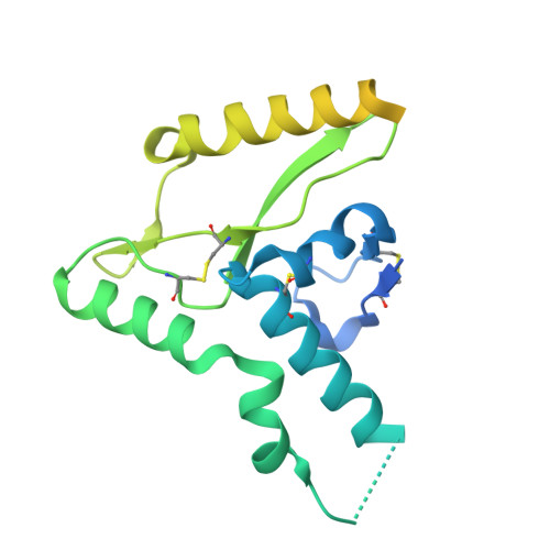
Metal-induced conformational changes in the Sabia virus spike complex.

Cohen-Dvashi, H.Katz, M.Diskin, R.

(2025) Nat Microbiol 10: 2221-2230

  • DOI: https://doi.org/10.1038/s41564-025-02075-8
  • Primary Citation of Related Structures:  
    9FYA, 9FYE, 9FYG

  • PubMed Abstract: 

    Haemorrhagic fever viruses from the Arenaviridae are a source of concern owing to their potential to cause lethal outbreaks and the lack of effective therapeutics. While structures of spike proteins from 'Old World' arenaviruses are available, the differences and similarities to 'New World' arenaviruses, such as the Sabiá virus, remain unclear owing to the lack of New World spike structures. Here we present the structure of the isolated spike complex from the Sabiá virus, which mediates viral attachment and entry to the host cells, using single-particle cryo-electron microscopy. We find two distinct conformational states of the spike, representing its native closed state at 2.6 Å resolution and an open state at 2.9 Å resolution that it assumes during cell entry. In addition, we show that the opening of the spike and subsequent cell entry are dependent on acidic pH and an unidentified metal ion. Our study suggests potential differences in the cell entry mechanisms of clade B arenaviruses compared with others in the Arenaviridae family.


  • Organizational Affiliation
    • Department of Chemical and Structural Biology, Weizmann Institute of Science, Rehovot, Israel.

Macromolecules
Find similar proteins by:  (by identity cutoff)  |  3D Structure
Entity ID: 1
MoleculeChains Sequence LengthOrganismDetailsImage
Glycoprotein G2A [auth a],
C [auth c],
D [auth b]
246Sabia virusMutation(s): 0 
Gene Names: GPCGP-C
UniProt
Find proteins for Q90037 (Sabia mammarenavirus (isolate Human/Brasil/SPH114202/1990))
Explore Q90037 
Go to UniProtKB:  Q90037
Entity Groups  
Sequence Clusters30% Identity50% Identity70% Identity90% Identity95% Identity100% Identity
UniProt GroupQ90037
Glycosylation
Glycosylation Sites: 4
Sequence Annotations
Expand
  • Reference Sequence
Find similar proteins by:  (by identity cutoff)  |  3D Structure
Entity ID: 2
MoleculeChains Sequence LengthOrganismDetailsImage
Glycoprotein G1B [auth A],
E [auth C],
F [auth B]
196Sabia virusMutation(s): 0 
Gene Names: GPCGP-C
UniProt
Find proteins for Q90037 (Sabia mammarenavirus (isolate Human/Brasil/SPH114202/1990))
Explore Q90037 
Go to UniProtKB:  Q90037
Entity Groups  
Sequence Clusters30% Identity50% Identity70% Identity90% Identity95% Identity100% Identity
UniProt GroupQ90037
Glycosylation
Glycosylation Sites: 7
Sequence Annotations
Expand
  • Reference Sequence
Oligosaccharides

Help

Entity ID: 3
MoleculeChains Length2D Diagram Glycosylation3D Interactions
2-acetamido-2-deoxy-beta-D-glucopyranose-(1-4)-2-acetamido-2-deoxy-beta-D-glucopyranose
G [auth D],
L [auth I],
N [auth K],
O [auth L],
T [auth Q],
G [auth D],
L [auth I],
N [auth K],
O [auth L],
T [auth Q],
Z [auth W]
2N-Glycosylation
Glycosylation Resources
GlyTouCan:  G42666HT
GlyCosmos:  G42666HT
GlyGen:  G42666HT
Entity ID: 4
MoleculeChains Length2D Diagram Glycosylation3D Interactions
alpha-D-mannopyranose-(1-3)-[alpha-D-mannopyranose-(1-6)]beta-D-mannopyranose-(1-4)-2-acetamido-2-deoxy-beta-D-glucopyranose-(1-4)-2-acetamido-2-deoxy-beta-D-glucopyranose
H [auth E],
J [auth G],
P [auth M],
R [auth O],
V [auth S],
H [auth E],
J [auth G],
P [auth M],
R [auth O],
V [auth S],
X [auth U]
5N-Glycosylation
Glycosylation Resources
GlyTouCan:  G22768VO
GlyCosmos:  G22768VO
GlyGen:  G22768VO
Entity ID: 5
MoleculeChains Length2D Diagram Glycosylation3D Interactions
beta-D-mannopyranose-(1-4)-2-acetamido-2-deoxy-beta-D-glucopyranose-(1-4)-2-acetamido-2-deoxy-beta-D-glucopyranose
AA [auth X],
I [auth F],
K [auth H],
M [auth J],
Q [auth N],
AA [auth X],
I [auth F],
K [auth H],
M [auth J],
Q [auth N],
S [auth P],
U [auth R],
W [auth T],
Y [auth V]
3N-Glycosylation
Glycosylation Resources
GlyTouCan:  G15407YE
GlyCosmos:  G15407YE
GlyGen:  G15407YE
Small Molecules
Ligands 2 Unique
IDChains Name / Formula / InChI Key2D Diagram3D Interactions
NAG
Query on NAG

Download Ideal Coordinates CCD File 
BA [auth a]
CA [auth a]
DA [auth a]
FA [auth A]
GA [auth c]
BA [auth a],
CA [auth a],
DA [auth a],
FA [auth A],
GA [auth c],
HA [auth c],
IA [auth c],
JA [auth b],
KA [auth b],
LA [auth b],
MA [auth C],
NA [auth B]
2-acetamido-2-deoxy-beta-D-glucopyranose
C8 H15 N O6
OVRNDRQMDRJTHS-FMDGEEDCSA-N
ZN
Query on ZN

Download Ideal Coordinates CCD File 
EA [auth a]ZINC ION
Zn
PTFCDOFLOPIGGS-UHFFFAOYSA-N
Experimental Data & Validation

Experimental Data

  • Method: ELECTRON MICROSCOPY
  • Resolution: 2.64 Å
  • Aggregation State: PARTICLE 
  • Reconstruction Method: SINGLE PARTICLE 
EM Software:
TaskSoftware PackageVersion
MODEL REFINEMENTPHENIX1.21.1_5286
RECONSTRUCTIONcryoSPARC4.5.3

Structure Validation

View Full Validation Report

Currently 9FYA does not have a validation slider image.


Entry History & Funding Information

Deposition Data


Funding OrganizationLocationGrant Number
Israel Science FoundationIsrael209/20

Revision History  (Full details and data files)

  • Version 1.0: 2025-06-11
    Type: Initial release
  • Version 1.1: 2025-08-13
    Changes: Data collection, Database references
  • Version 1.2: 2025-09-17
    Changes: Data collection, Database references