9G0T | pdb_00009g0t

Xenopus tropicalis undecorated microtubule - 15 protofilament, 3-start helix


Experimental Data Snapshot

  • Method: ELECTRON MICROSCOPY
  • Resolution: 3.50 Å
  • Aggregation State: FILAMENT 
  • Reconstruction Method: SINGLE PARTICLE 

Starting Model: other
View more details

wwPDB Validation   3D Report Full Report


This is version 1.2 of the entry. See complete history


Literature

Mechanistic basis of temperature adaptation in microtubule dynamics across frog species.

Troman, L.de Gaulejac, E.Biswas, A.Stiens, J.Kuropka, B.Moores, C.A.Reber, S.

(2025) Curr Biol 35: 612-628.e6

  • DOI: https://doi.org/10.1016/j.cub.2024.12.022
  • Primary Citation of Related Structures:  
    9FVJ, 9G0O, 9G0P, 9G0Q, 9G0R, 9G0S, 9G0T

  • PubMed Abstract: 

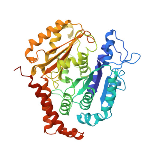
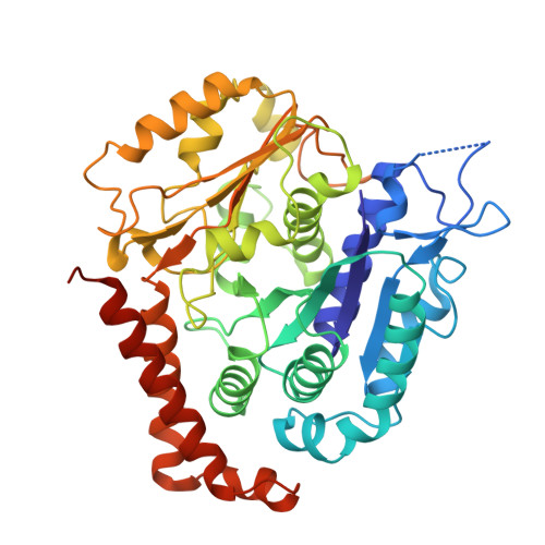
    Cellular processes are remarkably effective across diverse temperature ranges, even with highly conserved proteins. In the context of the microtubule cytoskeleton, which is critically involved in a wide range of cellular activities, this is particularly striking, as tubulin is one of the most conserved proteins while microtubule dynamic instability is highly temperature sensitive. Here, we leverage the diversity of natural tubulin variants from three closely related frog species that live at different temperatures. We determine the microtubule structure across all three species at between 3.0 and 3.6 Å resolution by cryo-electron microscopy and find small differences at the β-tubulin lateral interactions. Using in vitro reconstitution assays and quantitative biochemistry, we show that tubulin's free energy scales inversely with temperature. The observed weakening of lateral contacts and the low apparent activation energy for tubulin incorporation provide an explanation for the overall stability and higher growth rates of microtubules in cold-adapted frog species. This study thus broadens our conceptual framework for understanding microtubule dynamics and provides insights into how conserved cellular processes are tailored to different ecological niches.


  • Organizational Affiliation

    Institute of Structural and Molecular Biology, Birkbeck, University of London, Malet Street, London WC1E 7HX, UK.


Macromolecules
Find similar proteins by:  (by identity cutoff)  |  3D Structure
Entity ID: 1
MoleculeChains Sequence LengthOrganismDetailsImage
Tubulin beta chain
A, B, C, D, E
A, B, C, D, E, F
445Xenopus tropicalisMutation(s): 0 
UniProt
Find proteins for P30883 (Xenopus laevis)
Explore P30883 
Go to UniProtKB:  P30883
Entity Groups  
Sequence Clusters30% Identity50% Identity70% Identity90% Identity95% Identity100% Identity
UniProt GroupP30883
Sequence Annotations
Expand
  • Reference Sequence
Find similar proteins by:  (by identity cutoff)  |  3D Structure
Entity ID: 2
MoleculeChains Sequence LengthOrganismDetailsImage
Tubulin alpha chain449Xenopus tropicalisMutation(s): 0 
UniProt
Find proteins for Q5EB23 (Xenopus tropicalis)
Explore Q5EB23 
Go to UniProtKB:  Q5EB23
Entity Groups  
Sequence Clusters30% Identity50% Identity70% Identity90% Identity95% Identity100% Identity
UniProt GroupQ5EB23
Sequence Annotations
Expand
  • Reference Sequence
Small Molecules
Ligands 3 Unique
IDChains Name / Formula / InChI Key2D Diagram3D Interactions
GTP (Subject of Investigation/LOI)
Query on GTP

Download Ideal Coordinates CCD File 
AA [auth e]
CA [auth f]
S [auth a]
U [auth b]
W [auth c]
AA [auth e],
CA [auth f],
S [auth a],
U [auth b],
W [auth c],
Y [auth d]
GUANOSINE-5'-TRIPHOSPHATE
C10 H16 N5 O14 P3
XKMLYUALXHKNFT-UUOKFMHZSA-N
GDP (Subject of Investigation/LOI)
Query on GDP

Download Ideal Coordinates CCD File 
M [auth A]
N [auth B]
O [auth C]
P [auth D]
Q [auth E]
M [auth A],
N [auth B],
O [auth C],
P [auth D],
Q [auth E],
R [auth F]
GUANOSINE-5'-DIPHOSPHATE
C10 H15 N5 O11 P2
QGWNDRXFNXRZMB-UUOKFMHZSA-N
MG (Subject of Investigation/LOI)
Query on MG

Download Ideal Coordinates CCD File 
BA [auth e]
DA [auth f]
T [auth a]
V [auth b]
X [auth c]
BA [auth e],
DA [auth f],
T [auth a],
V [auth b],
X [auth c],
Z [auth d]
MAGNESIUM ION
Mg
JLVVSXFLKOJNIY-UHFFFAOYSA-N
Experimental Data & Validation

Experimental Data

  • Method: ELECTRON MICROSCOPY
  • Resolution: 3.50 Å
  • Aggregation State: FILAMENT 
  • Reconstruction Method: SINGLE PARTICLE 
EM Software:
TaskSoftware PackageVersion
RECONSTRUCTIONRELION3.1
MODEL REFINEMENTPHENIX

Structure Validation

View Full Validation Report


Entry History & Funding Information

Deposition Data


Funding OrganizationLocationGrant Number
Medical Research Council (MRC, United Kingdom)United KingdomMR/R000352/1
Medical Research Council (MRC, United Kingdom)United KingdomMR/Y000633/1
Wellcome TrustUnited Kingdom202679/Z/16/Z
Wellcome TrustUnited Kingdom206166/Z/17/Z

Revision History  (Full details and data files)

  • Version 1.0: 2025-01-15
    Type: Initial release
  • Version 1.1: 2025-01-22
    Changes: Data collection, Database references
  • Version 1.2: 2025-02-12
    Changes: Data collection, Database references