9GFD | pdb_00009gfd

Crystal structure of ASO binding Fab fragment with ASO139


Experimental Data Snapshot

  • Method: X-RAY DIFFRACTION
  • Resolution: 1.43 Å
  • R-Value Free: 
    0.205 (Depositor), 0.205 (DCC) 
  • R-Value Work: 
    0.183 (Depositor), 0.183 (DCC) 
  • R-Value Observed: 
    0.184 (Depositor) 

Starting Model: experimental
View more details

wwPDB Validation   3D Report Full Report


Ligand Structure Quality Assessment 


This is version 1.0 of the entry. See complete history


Literature

Improved Targeted Delivery of Antisense Oligonucleotide Conjugates with the Antibody Mask

Hsia, H.-E.Zanini, C.Simonneau, C.Fraidling, J.Kraft, T.Mayer, K.Sommer, A.Indlekofer, A.Wirth, T.Benz, J.Geroges, G.Langer, M.L.Gassner, C.Larraillet, V.Manso, M.Ravn, J.Hofer, K.Emrich, T.Niewoehner, J.Schumacher, F.Brinkmann, U.

To be published.

Macromolecules
Find similar proteins by:  (by identity cutoff)  |  3D Structure
Entity ID: 1
MoleculeChains Sequence LengthOrganismDetailsImage
Fab fragment heavy chainA [auth H]226Homo sapiensMutation(s): 0 
Entity Groups  
Sequence Clusters30% Identity50% Identity70% Identity90% Identity95% Identity100% Identity
Sequence Annotations
Expand
  • Reference Sequence
Find similar proteins by:  (by identity cutoff)  |  3D Structure
Entity ID: 2
MoleculeChains Sequence LengthOrganismDetailsImage
Fab Fragment light chainB [auth L]219Homo sapiensMutation(s): 0 
Entity Groups  
Sequence Clusters30% Identity50% Identity70% Identity90% Identity95% Identity100% Identity
Sequence Annotations
Expand
  • Reference Sequence
Small Molecules
Ligands 3 Unique
IDChains Name / Formula / InChI Key2D Diagram3D Interactions
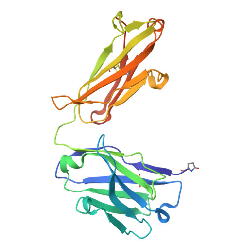
A1IKZ (Subject of Investigation/LOI)
Query on A1IKZ

Download Ideal Coordinates CCD File 
J [auth L]1-[(1~{S},3~{R},4~{R},7~{S})-7-[[(1~{R},3~{R},4~{R},7~{S})-7-[[(1~{R},3~{R},4~{R},7~{S})-3-(6-aminopurin-9-yl)-7-[[(2~{R},3~{S},5~{R})-5-(4-azanyl-2-oxidanylidene-pyrimidin-1-yl)-3-oxidanyl-oxolan-2-yl]methoxy-sulfanyl-phosphoryl]oxy-2,5-dioxabicyclo[2.2.1]heptan-1-yl]methoxy-sulfanyl-phosphoryl]oxy-3-(4-azanyl-5-methyl-2-oxidanylidene-pyrimidin-1-yl)-2,5-dioxabicyclo[2.2.1]heptan-1-yl]methoxy-sulfanyl-phosphoryl]oxy-1-(hydroxymethyl)-2,5-dioxabicyclo[2.2.1]heptan-3-yl]-5-methyl-pyrimidine-2,4-dione
C42 H52 N13 O22 P3 S3
YGGSNWRUJSQEEQ-LYYFKHMFSA-N
HIS
Query on HIS

Download Ideal Coordinates CCD File 
G [auth L]HISTIDINE
C6 H10 N3 O2
HNDVDQJCIGZPNO-YFKPBYRVSA-O
EDO
Query on EDO

Download Ideal Coordinates CCD File 
C [auth H]
D [auth H]
E [auth H]
F [auth L]
H [auth L]
C [auth H],
D [auth H],
E [auth H],
F [auth L],
H [auth L],
I [auth L]
1,2-ETHANEDIOL
C2 H6 O2
LYCAIKOWRPUZTN-UHFFFAOYSA-N
Modified Residues  1 Unique
IDChains TypeFormula2D DiagramParent
PCA
Query on PCA
A [auth H]L-PEPTIDE LINKINGC5 H7 N O3GLN
Experimental Data & Validation

Experimental Data

  • Method: X-RAY DIFFRACTION
  • Resolution: 1.43 Å
  • R-Value Free:  0.205 (Depositor), 0.205 (DCC) 
  • R-Value Work:  0.183 (Depositor), 0.183 (DCC) 
  • R-Value Observed: 0.184 (Depositor) 
Space Group: C 1 2 1
Unit Cell:
Length ( Å )Angle ( ˚ )
a = 125.824α = 90
b = 58.044β = 94.344
c = 62.472γ = 90
Software Package:
Software NamePurpose
PHENIXrefinement
XDSdata reduction
SADABSdata scaling
PHASERphasing

Structure Validation

View Full Validation Report


Ligand Structure Quality Assessment 


Entry History & Funding Information

Deposition Data


Funding OrganizationLocationGrant Number
Not funded--

Revision History  (Full details and data files)

  • Version 1.0: 2025-08-20
    Type: Initial release