9GU8 | pdb_00009gu8

NCS-1 bound to a FDA ligand 4


Experimental Data Snapshot

  • Method: X-RAY DIFFRACTION
  • Resolution: 1.67 Å
  • R-Value Free: 
    0.234 (Depositor), 0.234 (DCC) 
  • R-Value Work: 
    0.205 (Depositor), 0.205 (DCC) 
  • R-Value Observed: 
    0.207 (Depositor) 

Starting Model: experimental
View more details

wwPDB Validation   3D Report Full Report


Ligand Structure Quality Assessment 


This is version 1.0 of the entry. See complete history


Literature

FDA Drug Repurposing Uncovers Modulators of Dopamine D 2 Receptor Localization via Disruption of the NCS-1 Interaction.

Munoz-Reyes, D.Aguado, L.Arroyo-Urea, S.Requena, C.Perez-Suarez, S.Sanchez-Yepes, S.Argerich, J.Miro-Rodriguez, C.Ulzurrun, E.Rodriguez-Martin, E.Garcia-Nafria, J.Campillo, N.E.Mansilla, A.Sanchez-Barrena, M.J.

(2025) J Med Chem 

  • DOI: https://doi.org/10.1021/acs.jmedchem.5c01626
  • Primary Citation of Related Structures:  
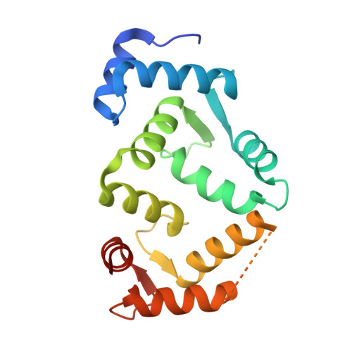
    9GTO, 9GU6, 9GU8

  • PubMed Abstract: 

    Dopamine D 2 receptor (D 2 R) regulates key aspects of motor control, cognition, and reward. Its function depends not only on ligand binding and signaling efficacy but also on the dynamic control of receptor localization at the cell surface. Neuronal calcium sensor 1 (NCS-1) interacts with D 2 R in a Ca 2+ -dependent manner. Using in vitro and cellular assays, we found that NCS-1 promotes D 2 R trafficking to the plasma membrane through active exocytosis while preserving canonical receptor pharmacology. A screen of FDA-approved drugs identified protein-protein interaction (PPI) modulators targeting the NCS-1/D 2 R interface. Azilsartan medoxomil, atorvastatin, and vilazodone disrupt this interaction, reducing D 2 R surface expression. Structural studies revealed that these compounds target NCS-1, overlap the D 2 R binding site, and perturb the dynamics of the regulatory helix H10. These findings reveal an unexploited intracellular mechanism to modulate D 2 R function via PPI modulation, offering a novel strategy to fine-tune dopaminergic tone beyond receptor blockade or direct agonism.


  • Organizational Affiliation
    • Department of Crystallography and Structural Biology, Institute of Physical-Chemistry "Blas Cabrera", CSIC, Serrano 119, 28006 Madrid, Spain.

Macromolecules
Find similar proteins by:  (by identity cutoff)  |  3D Structure
Entity ID: 1
MoleculeChains Sequence LengthOrganismDetailsImage
Neuronal calcium sensor 1A [auth B]177Homo sapiensMutation(s): 0 
Gene Names: NCS1FLUPFREQ
UniProt & NIH Common Fund Data Resources
Find proteins for P62166 (Homo sapiens)
Explore P62166 
Go to UniProtKB:  P62166
PHAROS:  P62166
GTEx:  ENSG00000107130 
Entity Groups  
Sequence Clusters30% Identity50% Identity70% Identity90% Identity95% Identity100% Identity
UniProt GroupP62166
Sequence Annotations
Expand
  • Reference Sequence
Small Molecules
Ligands 5 Unique
IDChains Name / Formula / InChI Key2D Diagram3D Interactions
YG7 (Subject of Investigation/LOI)
Query on YG7

Download Ideal Coordinates CCD File 
E [auth B]5-{4-[4-(5-cyano-1H-indol-3-yl)butyl]piperazin-1-yl}-1-benzofuran-2-carboxamide
C26 H27 N5 O2
SGEGOXDYSFKCPT-UHFFFAOYSA-N
1PE
Query on 1PE

Download Ideal Coordinates CCD File 
F [auth B],
G [auth B]
PENTAETHYLENE GLYCOL
C10 H22 O6
JLFNLZLINWHATN-UHFFFAOYSA-N
GOL
Query on GOL

Download Ideal Coordinates CCD File 
H [auth B],
I [auth B]
GLYCEROL
C3 H8 O3
PEDCQBHIVMGVHV-UHFFFAOYSA-N
CA
Query on CA

Download Ideal Coordinates CCD File 
B,
C [auth B],
D [auth B]
CALCIUM ION
Ca
BHPQYMZQTOCNFJ-UHFFFAOYSA-N
NA
Query on NA

Download Ideal Coordinates CCD File 
J [auth B],
K [auth B]
SODIUM ION
Na
FKNQFGJONOIPTF-UHFFFAOYSA-N
Experimental Data & Validation

Experimental Data

  • Method: X-RAY DIFFRACTION
  • Resolution: 1.67 Å
  • R-Value Free:  0.234 (Depositor), 0.234 (DCC) 
  • R-Value Work:  0.205 (Depositor), 0.205 (DCC) 
  • R-Value Observed: 0.207 (Depositor) 
Space Group: C 2 2 21
Unit Cell:
Length ( Å )Angle ( ˚ )
a = 44.608α = 90
b = 102.912β = 90
c = 76.718γ = 90
Software Package:
Software NamePurpose
autoPROCdata reduction
Aimlessdata scaling
PHASERphasing
PHENIXrefinement
Cootmodel building

Structure Validation

View Full Validation Report


Ligand Structure Quality Assessment 


Entry History & Funding Information

Deposition Data


Funding OrganizationLocationGrant Number
Ministerio de Ciencia e Innovacion (MCIN)SpainPID2022-137331OB-C31

Revision History  (Full details and data files)

  • Version 1.0: 2025-11-19
    Type: Initial release