9HKF | pdb_00009hkf

X-Ray crystal structure of a photoswitchable HaloTag bound to JF635


Experimental Data Snapshot

  • Method: X-RAY DIFFRACTION
  • Resolution: 2.40 Å
  • R-Value Free: 
    0.235 (Depositor), 0.235 (DCC) 
  • R-Value Work: 
    0.185 (Depositor), 0.189 (DCC) 
  • R-Value Observed: 
    0.188 (Depositor) 

Starting Model: experimental
View more details

wwPDB Validation   3D Report Full Report


Ligand Structure Quality Assessment 


This is version 1.0 of the entry. See complete history


Literature

A Photoswitchable HaloTag for Spatiotemporal Control of Fluorescence in Living Cells.

Walterspiel, F.Ugarte-Uribe, B.Weidenhausen, J.Vincent, M.Narayanasamy, K.K.Dimitriadi, A.Khan, A.U.M.Fritsch, M.Muller, C.W.Zimmermann, T.Deo, C.

(2025) Angew Chem Int Ed Engl : e202424955-e202424955

  • DOI: https://doi.org/10.1002/anie.202424955
  • Primary Citation of Related Structures:  
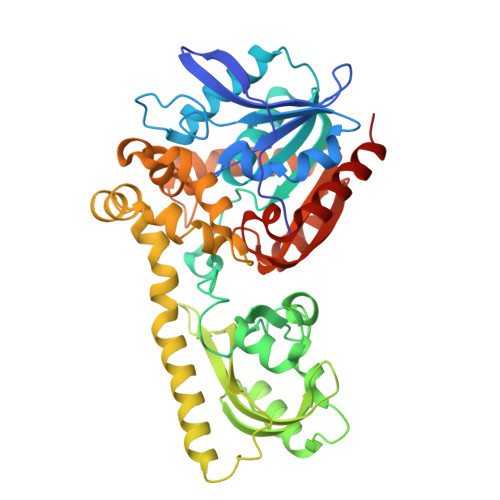
    9HKF

  • PubMed Abstract: 

    Photosensitive fluorophores, whose emission can be controlled using light, are essential for advanced biological imaging, enabling precise spatiotemporal tracking of molecular features and facilitating super-resolution microscopy techniques. Although irreversibly photoactivatable fluorophores are well established, reversible reporters that can be reactivated multiple times remain scarce, and only a few have been applied in living cells using generalizable protein labeling methods. To address these limitations, we introduce chemigenetic photoswitchable fluorophores, leveraging the self-labeling HaloTag protein with fluorogenic rhodamine dye ligands. By incorporating a light-responsive protein domain into HaloTag, we engineer a tunable, photoswitchable HaloTag (psHaloTag), which can reversibly modulate the fluorescence of a bound dye-ligand via a light-induced conformational change. Our best performing psHaloTag variants show excellent performance in living cells, with large, reversible, deep-red fluorescence turn-on upon 450 nm illumination across various biomolecular targets and SMLM compatibility. Together, this work establishes the chemigenetic approach as a versatile platform for the design of photoswitchable reporters, tunable through both genetic and synthetic modifications, with promising applications for dynamic imaging.


  • Organizational Affiliation
    • European Molecular Biology Laboratory, Meyerhofstraße 1, 69117, Heidelberg, Germany.

Macromolecules
Find similar proteins by:  (by identity cutoff)  |  3D Structure
Entity ID: 1
MoleculeChains Sequence LengthOrganismDetailsImage
Haloalkane dehalogenase,non-specific serine/threonine protein kinaseA [auth B],
B [auth A]
439Rhodococcus rhodochrousMutation(s): 24 
Gene Names: dhaA
EC: 3.8.1.5 (PDB Primary Data), 2.7.11.1 (PDB Primary Data)
UniProt
Find proteins for A0A453KFI0 (Aegilops tauschii subsp. strangulata)
Explore A0A453KFI0 
Go to UniProtKB:  A0A453KFI0
Find proteins for P0A3G2 (Rhodococcus rhodochrous)
Explore P0A3G2 
Go to UniProtKB:  P0A3G2
Entity Groups  
Sequence Clusters30% Identity50% Identity70% Identity90% Identity95% Identity100% Identity
UniProt GroupsP0A3G2A0A453KFI0
Sequence Annotations
Expand
  • Reference Sequence
Small Molecules
Ligands 4 Unique
IDChains Name / Formula / InChI Key2D Diagram3D Interactions
PUJ (Subject of Investigation/LOI)
Query on PUJ

Download Ideal Coordinates CCD File 
E [auth B],
L [auth A]
(1E,3S)-1-{10-[2-carboxy-5-({2-[2-(hexyloxy)ethoxy]ethyl}carbamoyl)phenyl]-7-(3-fluoroazetidin-1-yl)-5,5-dimethyldibenz o[b,e]silin-3(5H)-ylidene}-3-fluoroazetidin-1-ium
C39 H48 F2 N3 O5 Si
WMFJHJNMBLRDFB-UHFFFAOYSA-O
FMN (Subject of Investigation/LOI)
Query on FMN

Download Ideal Coordinates CCD File 
D [auth B],
K [auth A]
FLAVIN MONONUCLEOTIDE
C17 H21 N4 O9 P
FVTCRASFADXXNN-SCRDCRAPSA-N
GOL
Query on GOL

Download Ideal Coordinates CCD File 
F [auth B]
G [auth B]
H [auth B]
I [auth B]
M [auth A]
F [auth B],
G [auth B],
H [auth B],
I [auth B],
M [auth A],
N [auth A],
O [auth A],
P [auth A],
Q [auth A],
R [auth A],
S [auth A]
GLYCEROL
C3 H8 O3
PEDCQBHIVMGVHV-UHFFFAOYSA-N
CL
Query on CL

Download Ideal Coordinates CCD File 
C [auth B],
J [auth A]
CHLORIDE ION
Cl
VEXZGXHMUGYJMC-UHFFFAOYSA-M
Experimental Data & Validation

Experimental Data

  • Method: X-RAY DIFFRACTION
  • Resolution: 2.40 Å
  • R-Value Free:  0.235 (Depositor), 0.235 (DCC) 
  • R-Value Work:  0.185 (Depositor), 0.189 (DCC) 
  • R-Value Observed: 0.188 (Depositor) 
Space Group: C 1 2 1
Unit Cell:
Length ( Å )Angle ( ˚ )
a = 167.737α = 90
b = 78.017β = 103.92
c = 81.807γ = 90
Software Package:
Software NamePurpose
XDSdata reduction
Aimlessdata scaling
PHASERphasing
PHENIXrefinement

Structure Validation

View Full Validation Report


Ligand Structure Quality Assessment 


Entry History & Funding Information

Deposition Data


Funding OrganizationLocationGrant Number
Chan Zuckerberg InitiativeUnited States2020-225346
Chan Zuckerberg InitiativeUnited States2024-337799

Revision History  (Full details and data files)

  • Version 1.0: 2025-11-05
    Type: Initial release