9HPM | pdb_00009hpm

E. coli beta-galactosidase labeled with Chromeo P503 dye purified using MISO from 1ug


Experimental Data Snapshot

  • Method: ELECTRON MICROSCOPY
  • Resolution: 2.20 Å
  • Aggregation State: PARTICLE 
  • Reconstruction Method: SINGLE PARTICLE 

wwPDB Validation   3D Report Full Report


This is version 1.1 of the entry. See complete history


Literature

MISO: microfluidic protein isolation enables single-particle cryo-EM structure determination from a single cell colony.

Eluru, G.De Gieter, S.Schenck, S.Stroobants, A.Shrestha, B.Erbel, P.Brunner, J.D.Efremov, R.G.

(2025) Nat Methods 

  • DOI: https://doi.org/10.1038/s41592-025-02894-x
  • Primary Citation of Related Structures:  
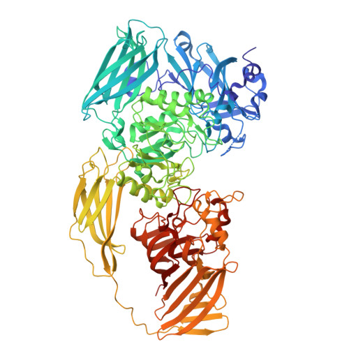
    9HPL, 9HPM, 9HQN, 9HQO, 9HQP

  • PubMed Abstract: 

    Single-particle cryogenic electron microscopy (cryo-EM) enables reconstruction of atomic-resolution 3D maps of proteins by visualizing thousands to millions of purified protein particles embedded in vitreous ice. This corresponds to picograms of purified protein, which can potentially be isolated from a few thousand cells. Hence, cryo-EM holds the potential of a very sensitive analytical method for delivering high-resolution protein structure as a readout. In practice, millions of times more starting biological material is required to prepare cryo-EM grids. Here we show that using a micro isolation (MISO) method, which combines microfluidics-based protein purification with cryo-EM grid preparation, cryo-EM structures of soluble bacterial and eukaryotic membrane proteins can be solved starting from less than 1 µg of a target protein and progressing from cells to cryo-EM grids within a few hours. This scales down the amount of starting biological material hundreds to thousands of times, opening possibilities for the structural characterization of hitherto inaccessible proteins.


  • Organizational Affiliation
    • Center for Structural Biology, Vlaams Instituut voor Biotechnologie, Brussels, Belgium.

Macromolecules
Find similar proteins by:  (by identity cutoff)  |  3D Structure
Entity ID: 1
MoleculeChains Sequence LengthOrganismDetailsImage
Beta-galactosidase
A, B, C, D
1,031Escherichia coli K-12Mutation(s): 0 
Gene Names: lacZb0344JW0335
EC: 3.2.1.23
UniProt
Find proteins for P00722 (Escherichia coli (strain K12))
Explore P00722 
Go to UniProtKB:  P00722
Entity Groups  
Sequence Clusters30% Identity50% Identity70% Identity90% Identity95% Identity100% Identity
UniProt GroupP00722
Sequence Annotations
Expand
  • Reference Sequence
Experimental Data & Validation

Experimental Data

  • Method: ELECTRON MICROSCOPY
  • Resolution: 2.20 Å
  • Aggregation State: PARTICLE 
  • Reconstruction Method: SINGLE PARTICLE 
EM Software:
TaskSoftware PackageVersion
MODEL REFINEMENTPHENIX1.20.1_4487

Structure Validation

View Full Validation Report


Entry History & Funding Information

Deposition Data


Funding OrganizationLocationGrant Number
European Research Council (ERC)European Union726436
Research Foundation - Flanders (FWO)BelgiumG0H5916N
Research Foundation - Flanders (FWO)BelgiumG054617N

Revision History  (Full details and data files)

  • Version 1.0: 2025-11-05
    Type: Initial release
  • Version 1.1: 2025-11-26
    Changes: Data collection, Database references