9I2H | pdb_00009i2h

A 3.3 angstrom cryo-EM structure of an engineered high-affinity human prothrombinase complex


Experimental Data Snapshot

  • Method: ELECTRON MICROSCOPY
  • Resolution: 3.28 Å
  • Aggregation State: PARTICLE 
  • Reconstruction Method: SINGLE PARTICLE 

wwPDB Validation   3D Report Full Report


This is version 1.0 of the entry. See complete history


Literature

A 3.3- angstrom cryo-EM structure of an engineered high-affinity human prothrombinase complex.

Ustok, F.I.Faille, A.Huntington, J.A.

(2025) Blood 

  • DOI: https://doi.org/10.1182/blood.2025031527
  • Primary Citation of Related Structures:  
    9I24, 9I2H

  • PubMed Abstract: 

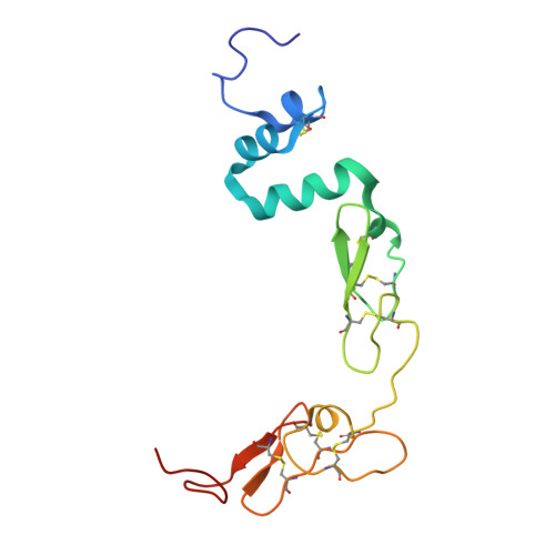
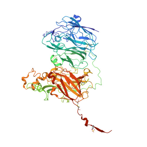
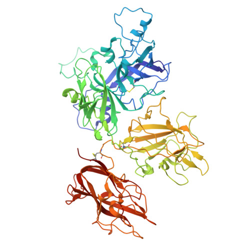
    Thrombin is generated from prothrombin through cleavage at two sites by the enzyme prothrombinase, composed of factor (f) Xa and fVa. The affinity of fXa for fVa is low, with assembly and function dependent on phospholipid (PL) membranes. Some snakes have evolved venom versions of fXa that bind to fVa with high affinity and efficiently activate prothrombin in the absence of PL. We created a similar high-affinity, PL-independent human prothrombinase with 17 mutations to human fXa (M17). The increase in affinity enabled cryogenic electron microscopy (cryo-EM) structure determination of M17-prothrombinase to a resolution of 3.3 Å. All protein domains were well resolved in the map, except for the Gla domain of fXa. The main contacts involve the serine protease and EGF2 domains of fXa and the A2 and A3 domains of fVa, resulting in the burying of a total surface area of 4,900 Å2. The map is of sufficient quality to resolve side chain interactions, including several key M17 mutations. To aid in the placement of the loop C-terminal to the A2 domain (a2-loop), we solved a high-resolution crystal structure of fXa in complex with a synthetic a2-peptide. The acidic a2-loop interacts with the basic heparin binding site of fXa, involving a conserved antiparallel b-strand interaction. The M17-prothrombinase structure is compatible with data from biochemical and mutagenesis research, and provides important new insights into the assembly and function of the prothrombinase complex.


  • Organizational Affiliation
    • University of Cambridge, Cambridge, United Kingdom.

Macromolecules
Find similar proteins by:  (by identity cutoff)  |  3D Structure
Entity ID: 1
MoleculeChains Sequence LengthOrganismDetailsImage
Activated factor Xa heavy chainA [auth H]254Homo sapiensMutation(s): 10 
Gene Names: F10
EC: 3.4.21.6
UniProt & NIH Common Fund Data Resources
Find proteins for P00742 (Homo sapiens)
Explore P00742 
Go to UniProtKB:  P00742
PHAROS:  P00742
GTEx:  ENSG00000126218 
Entity Groups  
Sequence Clusters30% Identity50% Identity70% Identity90% Identity95% Identity100% Identity
UniProt GroupP00742
Sequence Annotations
Expand
  • Reference Sequence
Find similar proteins by:  (by identity cutoff)  |  3D Structure
Entity ID: 2
MoleculeChains Sequence LengthOrganismDetailsImage
Factor X light chainB [auth L]139Homo sapiensMutation(s): 7 
Gene Names: F10
EC: 3.4.21.6
UniProt & NIH Common Fund Data Resources
Find proteins for P00742 (Homo sapiens)
Explore P00742 
Go to UniProtKB:  P00742
PHAROS:  P00742
GTEx:  ENSG00000126218 
Entity Groups  
Sequence Clusters30% Identity50% Identity70% Identity90% Identity95% Identity100% Identity
UniProt GroupP00742
Sequence Annotations
Expand
  • Reference Sequence
Find similar proteins by:  (by identity cutoff)  |  3D Structure
Entity ID: 3
MoleculeChains Sequence LengthOrganismDetailsImage
Coagulation factor V heavy chainC [auth A]709Homo sapiensMutation(s): 0 
Gene Names: F5
UniProt & NIH Common Fund Data Resources
Find proteins for P12259 (Homo sapiens)
Explore P12259 
Go to UniProtKB:  P12259
PHAROS:  P12259
GTEx:  ENSG00000198734 
Entity Groups  
Sequence Clusters30% Identity50% Identity70% Identity90% Identity95% Identity100% Identity
UniProt GroupP12259
Glycosylation
Glycosylation Sites: 2Go to GlyGen: P12259-1
Sequence Annotations
Expand
  • Reference Sequence
Find similar proteins by:  (by identity cutoff)  |  3D Structure
Entity ID: 4
MoleculeChains Sequence LengthOrganismDetailsImage
Coagulation factor V light chainD [auth B]651Homo sapiensMutation(s): 0 
Gene Names: F5
UniProt & NIH Common Fund Data Resources
Find proteins for P12259 (Homo sapiens)
Explore P12259 
Go to UniProtKB:  P12259
PHAROS:  P12259
GTEx:  ENSG00000198734 
Entity Groups  
Sequence Clusters30% Identity50% Identity70% Identity90% Identity95% Identity100% Identity
UniProt GroupP12259
Glycosylation
Glycosylation Sites: 2Go to GlyGen: P12259-1
Sequence Annotations
Expand
  • Reference Sequence
Oligosaccharides

Help

Entity ID: 5
MoleculeChains Length2D Diagram Glycosylation3D Interactions
alpha-D-mannopyranose-(1-3)-[alpha-D-mannopyranose-(1-6)]beta-D-mannopyranose-(1-4)-2-acetamido-2-deoxy-beta-D-glucopyranose-(1-4)-[alpha-L-fucopyranose-(1-6)]2-acetamido-2-deoxy-beta-D-glucopyranoseE [auth X]6N-Glycosylation
Entity ID: 6
MoleculeChains Length2D Diagram Glycosylation3D Interactions
alpha-D-mannopyranose-(1-3)-[alpha-D-mannopyranose-(1-6)]beta-D-mannopyranose-(1-4)-2-acetamido-2-deoxy-beta-D-glucopyranose-(1-4)-2-acetamido-2-deoxy-beta-D-glucopyranoseF [auth C]5N-Glycosylation
Entity ID: 7
MoleculeChains Length2D Diagram Glycosylation3D Interactions
alpha-D-mannopyranose-(1-6)-alpha-D-mannopyranose-(1-3)-[alpha-D-mannopyranose-(1-6)-alpha-D-mannopyranose-(1-6)]beta-D-mannopyranose-(1-4)-2-acetamido-2-deoxy-beta-D-glucopyranose-(1-4)-2-acetamido-2-deoxy-beta-D-glucopyranoseG [auth D]7N-Glycosylation
Entity ID: 8
MoleculeChains Length2D Diagram Glycosylation3D Interactions
alpha-L-fucopyranose-(1-6)-2-acetamido-2-deoxy-beta-D-glucopyranoseH [auth E]2N-Glycosylation
Small Molecules
Ligands 4 Unique
IDChains Name / Formula / InChI Key2D Diagram3D Interactions
0GJ
Query on 0GJ

Download Ideal Coordinates CCD File 
K [auth H]L-alpha-glutamyl-N-{(1S)-4-{[amino(iminio)methyl]amino}-1-[(1S)-2-chloro-1-hydroxyethyl]butyl}glycinamide
C14 H28 Cl N6 O5
XELWNHKFCNMWQO-LPEHRKFASA-O
CU
Query on CU

Download Ideal Coordinates CCD File 
M [auth B]COPPER (II) ION
Cu
JPVYNHNXODAKFH-UHFFFAOYSA-N
CA
Query on CA

Download Ideal Coordinates CCD File 
I [auth H],
L [auth A]
CALCIUM ION
Ca
BHPQYMZQTOCNFJ-UHFFFAOYSA-N
NA
Query on NA

Download Ideal Coordinates CCD File 
J [auth H]SODIUM ION
Na
FKNQFGJONOIPTF-UHFFFAOYSA-N
Modified Residues  1 Unique
IDChains TypeFormula2D DiagramParent
TYS
Query on TYS
C [auth A]L-PEPTIDE LINKINGC9 H11 N O6 STYR
Biologically Interesting Molecules (External Reference) 1 Unique
Experimental Data & Validation

Experimental Data

  • Method: ELECTRON MICROSCOPY
  • Resolution: 3.28 Å
  • Aggregation State: PARTICLE 
  • Reconstruction Method: SINGLE PARTICLE 
EM Software:
TaskSoftware PackageVersion
MODEL REFINEMENTPHENIX1.21.1_5286

Structure Validation

View Full Validation Report


Entry History & Funding Information

Deposition Data


Funding OrganizationLocationGrant Number
British Heart FoundationUnited KingdomRG/16/9/32391
British Heart FoundationUnited KingdomPG/24/11721

Revision History  (Full details and data files)

  • Version 1.0: 2025-11-26
    Type: Initial release