9I50 | pdb_00009i50

Crystal structure of feruloyl esterase from Fusarium oxysporum G122S variant


Experimental Data Snapshot

  • Method: X-RAY DIFFRACTION
  • Resolution: 1.90 Å
  • R-Value Free: 
    0.197 (Depositor), 0.200 (DCC) 
  • R-Value Work: 
    0.162 (Depositor), 0.165 (DCC) 

Starting Model: experimental
View more details

wwPDB Validation   3D Report Full Report


Ligand Structure Quality Assessment 


This is version 1.0 of the entry. See complete history


Literature

Structural insights into an engineered feruloyl esterase with improved MHET degrading properties.

Karampa, P.Makryniotis, K.Sousani, T.I.Topakas, E.Daskalakis, V.Dimarogona, M.

(2026) FEBS Lett 

  • DOI: https://doi.org/10.1002/1873-3468.70322
  • Primary Citation Related Structures: 
    9HUN, 9I50

  • PubMed Abstract: 

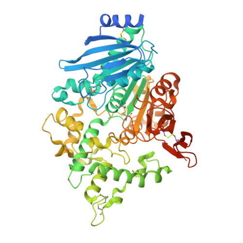
    Mono(2-hydroxyethyl) terephthalate (MHET) esterases (MHETases) are enzymes implicated in polyethylene terephthalate (PET) biodegradation. The present study elucidates the structural determinants that result in increased MHET degradation by a feruloyl esterase, which has been engineered to resemble the MHETase active site. The crystal structures of the variant in apo- and benzoic acid bound states reveal the changes induced by the introduced mutation, specifically the formation of a hydrogen bond and a trans to cis isomerization of a peptide bond in the vicinity of the catalytic site. Molecular dynamics simulations demonstrate the stabilization of the loop harboring the engineered residue, as well as an expansion of the substrate binding cleft, which would facilitate accommodation of a broader variety of substrates, indicative of a promiscuous biocatalyst.


  • Organizational Affiliation
    • Laboratory of Structural Biology and Biotechnology, Department of Chemical Engineering, University of Patras, Greece.

Macromolecules
Find similar proteins by:  (by identity cutoff)  |  3D Structure
Entity ID: 1
MoleculeChains Sequence LengthOrganismDetailsImage
Carboxylic ester hydrolase
A, B
563Fusarium oxysporumMutation(s): 1 
Gene Names: faeC
EC: 3.1.1
UniProt
Find proteins for A0A1D3S5H0 (Fusarium oxysporum)
Explore A0A1D3S5H0 
Go to UniProtKB:  A0A1D3S5H0
Entity Groups  
Sequence Clusters30% Identity50% Identity70% Identity90% Identity95% Identity100% Identity
UniProt GroupA0A1D3S5H0
Sequence Annotations
Expand
  • Reference Sequence
Oligosaccharides

Help

Entity ID: 2
MoleculeChains Length2D Diagram Glycosylation3D Interactions
beta-D-mannopyranose-(1-4)-2-acetamido-2-deoxy-beta-D-glucopyranose-(1-4)-2-acetamido-2-deoxy-beta-D-glucopyranose
C
3N/A
Entity ID: 3
MoleculeChains Length2D Diagram Glycosylation3D Interactions
alpha-D-mannopyranose-(1-2)-alpha-D-mannopyranose-(1-6)-alpha-D-mannopyranose-(1-6)-beta-D-mannopyranose-(1-4)-2-acetamido-2-deoxy-beta-D-glucopyranose-(1-4)-2-acetamido-2-deoxy-beta-D-glucopyranose
D
6N/A
Entity ID: 4
MoleculeChains Length2D Diagram Glycosylation3D Interactions
alpha-D-mannopyranose-(1-2)-alpha-D-mannopyranose-(1-6)-[alpha-D-mannopyranose-(1-3)]alpha-D-mannopyranose-(1-6)-[alpha-D-mannopyranose-(1-2)-alpha-D-mannopyranose-(1-3)]beta-D-mannopyranose-(1-4)-2-acetamido-2-deoxy-beta-D-glucopyranose-(1-4)-2-acetamido-2-deoxy-beta-D-glucopyranose
E
9N/A
Entity ID: 5
MoleculeChains Length2D Diagram Glycosylation3D Interactions
alpha-D-mannopyranose-(1-2)-alpha-D-mannopyranose-(1-2)-alpha-D-mannopyranose-(1-3)-[alpha-D-mannopyranose-(1-2)-alpha-D-mannopyranose-(1-6)-[alpha-D-mannopyranose-(1-3)]alpha-D-mannopyranose-(1-6)]beta-D-mannopyranose-(1-4)-2-acetamido-2-deoxy-beta-D-glucopyranose-(1-4)-2-acetamido-2-deoxy-beta-D-glucopyranose
F, G
10N/A
Small Molecules
Ligands 5 Unique
IDChains Name / Formula / InChI Key2D Diagram3D Interactions
NAG
Query on NAG

Download Ideal Coordinates CCD File 
H [auth A],
O [auth A],
R [auth B],
Y [auth B],
Z [auth B]
2-acetamido-2-deoxy-beta-D-glucopyranose
C8 H15 N O6
OVRNDRQMDRJTHS-FMDGEEDCSA-N
PGE
Query on PGE

Download Ideal Coordinates CCD File 
N [auth A]TRIETHYLENE GLYCOL
C6 H14 O4
ZIBGPFATKBEMQZ-UHFFFAOYSA-N
PEG
Query on PEG

Download Ideal Coordinates CCD File 
BA [auth B]
CA [auth B]
DA [auth B]
I [auth A]
J [auth A]
BA [auth B],
CA [auth B],
DA [auth B],
I [auth A],
J [auth A],
L [auth A],
Q [auth A],
S [auth B]
DI(HYDROXYETHYL)ETHER
C4 H10 O3
MTHSVFCYNBDYFN-UHFFFAOYSA-N
EDO
Query on EDO

Download Ideal Coordinates CCD File 
K [auth A]
M [auth A]
T [auth B]
U [auth B]
V [auth B]
K [auth A],
M [auth A],
T [auth B],
U [auth B],
V [auth B],
W [auth B],
X [auth B]
1,2-ETHANEDIOL
C2 H6 O2
LYCAIKOWRPUZTN-UHFFFAOYSA-N
CA
Query on CA

Download Ideal Coordinates CCD File 
AA [auth B],
P [auth A]
CALCIUM ION
Ca
BHPQYMZQTOCNFJ-UHFFFAOYSA-N
Experimental Data & Validation

Experimental Data

  • Method: X-RAY DIFFRACTION
  • Resolution: 1.90 Å
  • R-Value Free:  0.197 (Depositor), 0.200 (DCC) 
  • R-Value Work:  0.162 (Depositor), 0.165 (DCC) 
Space Group: P 1 21 1
Unit Cell:
Length ( Å )Angle ( ˚ )
a = 68.561α = 90
b = 89.261β = 101.725
c = 116.484γ = 90
Software Package:
Software NamePurpose
REFMACrefinement
PHASERphasing
Aimlessdata scaling
XDSdata reduction

Structure Validation

View Full Validation Report


Ligand Structure Quality Assessment 


Entry History & Funding Information

Deposition Data


Funding OrganizationLocationGrant Number
Hellenic Foundation for Research and Innovation (HFRI)Greece03061

Revision History  (Full details and data files)

  • Version 1.0: 2026-04-01
    Type: Initial release