


Rewiring Catalytic Craters: A Path for Engineering beta-Glucosidases to Improve Glucose Tolerance
Suryawanshi, A.B., Bedi, R.K., Pawar, M., Noronha, S., Bhaumik, P.(2025) bioRxiv
Experimental Data Snapshot
Starting Model: experimental
View more details
wwPDB Validation 3D Report Full Report

(2025) bioRxiv
Entity ID: 1 | |||||
|---|---|---|---|---|---|
| Molecule | Chains | Sequence Length | Organism | Details | Image |
| Beta-glucosidase | 477 | soil metagenome | Mutation(s): 1 EC: 3.2.1.21 | 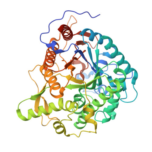 | |
Entity Groups | |||||
| Sequence Clusters | 30% Identity50% Identity70% Identity90% Identity95% Identity100% Identity | ||||
Sequence AnnotationsExpand | |||||
| |||||
| Ligands 2 Unique | |||||
|---|---|---|---|---|---|
| ID | Chains | Name / Formula / InChI Key | 2D Diagram | 3D Interactions | |
| PO4 Query on PO4 | C [auth A] | PHOSPHATE ION O4 P NBIIXXVUZAFLBC-UHFFFAOYSA-K |  | ||
| CL Query on CL | B [auth A] | CHLORIDE ION Cl VEXZGXHMUGYJMC-UHFFFAOYSA-M |  | ||
| Length ( Å ) | Angle ( ˚ ) |
|---|---|
| a = 51.88 | α = 90 |
| b = 66.27 | β = 90 |
| c = 138.25 | γ = 90 |
| Software Name | Purpose |
|---|---|
| REFMAC | refinement |
| XSCALE | data scaling |
| XDS | data reduction |
| PHASER | phasing |
| Funding Organization | Location | Grant Number |
|---|---|---|
| Department of Biotechnology (DBT, India) | India | BT/PR41982/PBD/26/822/2021 |


