9KUG | pdb_00009kug

Structure of EP67 bound to human C5aR1 in complex with Go


Experimental Data Snapshot

  • Method: ELECTRON MICROSCOPY
  • Resolution: 3.07 Å
  • Aggregation State: PARTICLE 
  • Reconstruction Method: SINGLE PARTICLE 

Starting Model: experimental
View more details

wwPDB Validation   3D Report Full Report


This is version 1.0 of the entry. See complete history


Literature

Molecular fingerprints of a convergent mechanism orchestrating diverse ligand recognition and species-specific pharmacology at the complement anaphylatoxin receptors.

Mishra, S.Yadav, M.K.Dalal, A.Ganguly, M.Yadav, R.Sawada, K.Tiwari, D.Roy, N.Banerjee, N.Fung, J.N.Marallag, J.Cui, C.S.Li, X.X.Lee, J.D.Dsouza, C.A.Saha, S.Sarma, P.Rawat, G.Zhu, H.Khant, H.A.Clark, R.J.Sano, F.K.Banerjee, R.Woodruff, T.M.Nureki, O.Gati, C.Shukla, A.K.

(2025) bioRxiv 

  • DOI: https://doi.org/10.1101/2025.05.26.656101
  • Primary Citation of Related Structures:  
    9KUG, 9KUT, 9KV6, 9KV8, 9KVP, 9KWG, 9KWX, 9KX6, 9KXS, 9KY2, 9KYU, 9KZ2, 9KZ8, 9KZK, 9L0H, 9UMJ, 9UMR, 9UMX

  • PubMed Abstract: 

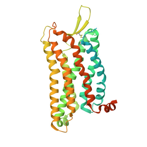
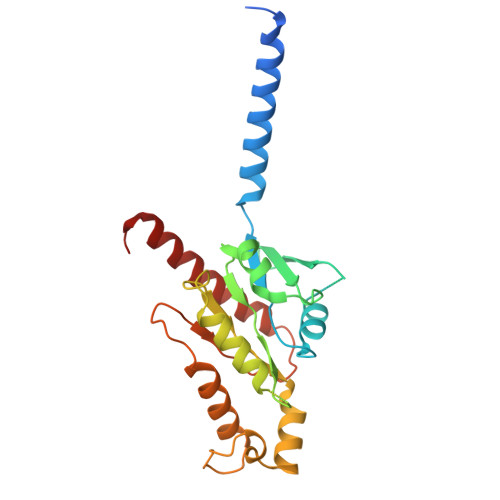
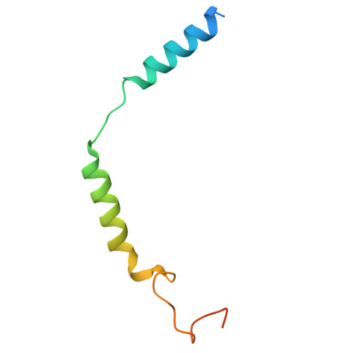
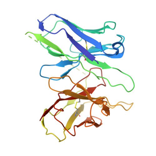
    Complement anaphylatoxin receptors (C3aR and C5aR1) are prototypical G protein-coupled receptors (GPCRs) playing crucial physiological roles in innate immunity by combating pathogenic infections and orchestrating inflammatory responses. They continue to be important therapeutic targets for multiple disorders including autoimmune diseases, acute and chronic inflammation, and allergy-related conditions. Recent structural coverage has provided important insights into their activation and signaling, however, confounding observations in the literature related to ligand efficacy and functional responses, especially in different model systems, present a major challenge for drug discovery efforts. Here, we systematically and comprehensively profile a broad set of natural and synthetic ligands at C3aR and C5aR1 and discover a previously unanticipated level of functional specialization in terms of species-specific pharmacology and receptor activation. Taking a lead from this, we determine seventeen cryo-EM structures of different ligand-receptor-G-protein complexes and uncover distinct orientation of agonists between the human and mouse receptors despite an overlapping positioning in the orthosteric binding pocket. Combined with extensive mutagenesis and functional assays, these structural snapshots allow us to decode and validate a convergent molecular mechanism involving a "Five-Point-Switch" in these receptors that orchestrates the recognition and efficacy of diverse agonists. We also identify species-specific differences at the level of phosphorylation patterns encoded in the carboxyl-terminus of these receptors and directly visualize their impact on βarr binding and activation using cryo-EM structures. Interestingly, we observe that βarrs engage with the mouse C5aR1 using a variation of previously discovered P-X-P-P phosphorylation motif via a "Sliding-Mechanism" and also exhibit distinct oligomeric state for the human vs. mouse receptors. Taken together, this study elucidates functional specialization at the complement anaphylatoxin receptors and underlying molecular mechanisms, offering a previously lacking framework with direct and immediate implications for the development of novel therapeutics.


  • Organizational Affiliation
    • Department of Biological Sciences and Bioengineering, Indian Institute of Technology, Kanpur 208016, India.

Macromolecules
Find similar proteins by:  (by identity cutoff)  |  3D Structure
Entity ID: 1
MoleculeChains Sequence LengthOrganismDetailsImage
Muscarinic acetylcholine receptor M4,C5a anaphylatoxin chemotactic receptor 1406Homo sapiensMutation(s): 0 
Gene Names: CHRM4C5AR1C5ARC5R1
UniProt & NIH Common Fund Data Resources
Find proteins for P08173 (Homo sapiens)
Explore P08173 
Go to UniProtKB:  P08173
PHAROS:  P08173
GTEx:  ENSG00000180720 
Find proteins for P21730 (Homo sapiens)
Explore P21730 
Go to UniProtKB:  P21730
PHAROS:  P21730
GTEx:  ENSG00000197405 
Entity Groups  
Sequence Clusters30% Identity50% Identity70% Identity90% Identity95% Identity100% Identity
UniProt GroupsP08173P21730
Sequence Annotations
Expand
  • Reference Sequence
Find similar proteins by:  (by identity cutoff)  |  3D Structure
Entity ID: 2
MoleculeChains Sequence LengthOrganismDetailsImage
Guanine nucleotide-binding protein G(o) subunit alpha250Homo sapiensMutation(s): 6 
Gene Names: GNAO1
EC: 3.6.5
UniProt & NIH Common Fund Data Resources
Find proteins for P09471 (Homo sapiens)
Explore P09471 
Go to UniProtKB:  P09471
PHAROS:  P09471
GTEx:  ENSG00000087258 
Entity Groups  
Sequence Clusters30% Identity50% Identity70% Identity90% Identity95% Identity100% Identity
UniProt GroupP09471
Sequence Annotations
Expand
  • Reference Sequence
Find similar proteins by:  (by identity cutoff)  |  3D Structure
Entity ID: 3
MoleculeChains Sequence LengthOrganismDetailsImage
Guanine nucleotide-binding protein G(I)/G(S)/G(T) subunit beta-1350Homo sapiensMutation(s): 0 
Gene Names: GNB1
UniProt & NIH Common Fund Data Resources
Find proteins for P62873 (Homo sapiens)
Explore P62873 
Go to UniProtKB:  P62873
PHAROS:  P62873
GTEx:  ENSG00000078369 
Entity Groups  
Sequence Clusters30% Identity50% Identity70% Identity90% Identity95% Identity100% Identity
UniProt GroupP62873
Sequence Annotations
Expand
  • Reference Sequence
Find similar proteins by:  (by identity cutoff)  |  3D Structure
Entity ID: 4
MoleculeChains Sequence LengthOrganismDetailsImage
Guanine nucleotide-binding protein G(I)/G(S)/G(O) subunit gamma-2D [auth G]71Homo sapiensMutation(s): 0 
Gene Names: GNG2
UniProt & NIH Common Fund Data Resources
Find proteins for P59768 (Homo sapiens)
Explore P59768 
Go to UniProtKB:  P59768
PHAROS:  P59768
GTEx:  ENSG00000186469 
Entity Groups  
Sequence Clusters30% Identity50% Identity70% Identity90% Identity95% Identity100% Identity
UniProt GroupP59768
Sequence Annotations
Expand
  • Reference Sequence
Find similar proteins by:  (by identity cutoff)  |  3D Structure
Entity ID: 5
MoleculeChains Sequence LengthOrganismDetailsImage
Antibody fragment - ScFv16E [auth H]248Mus musculusMutation(s): 0 
Entity Groups  
Sequence Clusters30% Identity50% Identity70% Identity90% Identity95% Identity100% Identity
Sequence Annotations
Expand
  • Reference Sequence

Find similar proteins by:  Sequence   |   3D Structure  

Entity ID: 6
MoleculeChains Sequence LengthOrganismDetailsImage
EP67F [auth D]10Homo sapiensMutation(s): 0 
Entity Groups  
Sequence Clusters30% Identity50% Identity70% Identity90% Identity95% Identity100% Identity
Sequence Annotations
Expand
  • Reference Sequence
Small Molecules
Modified Residues  1 Unique
IDChains TypeFormula2D DiagramParent
MLE
Query on MLE
F [auth D]L-PEPTIDE LINKINGC7 H15 N O2LEU
Experimental Data & Validation

Experimental Data

  • Method: ELECTRON MICROSCOPY
  • Resolution: 3.07 Å
  • Aggregation State: PARTICLE 
  • Reconstruction Method: SINGLE PARTICLE 
EM Software:
TaskSoftware PackageVersion
RECONSTRUCTIONcryoSPARC4.5.3
MODEL REFINEMENTPHENIX

Structure Validation

View Full Validation Report


Entry History & Funding Information

Deposition Data


Funding OrganizationLocationGrant Number
Wellcome TrustUnited KingdomIA/S/20/1/504916
Science and Engineering Research Board (SERB)IndiaIPA/2020/000405
Science and Engineering Research Board (SERB)IndiaCRG/2022/002646

Revision History  (Full details and data files)

  • Version 1.0: 2025-11-26
    Type: Initial release