9OMM | pdb_00009omm

WrtF fucosyltransferase - tetrasaccharide product


Experimental Data Snapshot

  • Method: X-RAY DIFFRACTION
  • Resolution: 2.25 Å
  • R-Value Free: 
    0.265 (Depositor), 0.267 (DCC) 
  • R-Value Work: 
    0.215 (Depositor), 0.225 (DCC) 
  • R-Value Observed: 
    0.220 (Depositor) 

Starting Model: experimental
View more details

wwPDB Validation   3D Report Full Report


Ligand Structure Quality Assessment 


This is version 1.1 of the entry. See complete history


Literature

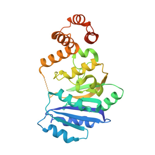
WrtF from Rhizobium tropici CIAT 899 is a GT-A fold fucosyltransferase that binds its donor nonproductively.

Forrester, T.J.B.Lin, S.Lowary, T.L.Kimber, M.S.

(2025) Proc Natl Acad Sci U S A 122: e2512460122-e2512460122

  • DOI: https://doi.org/10.1073/pnas.2512460122
  • Primary Citation of Related Structures:  
    9OMH, 9OMI, 9OMJ, 9OMK, 9OML, 9OMM, 9OMN

  • PubMed Abstract: 

    l-Fucose, a 6-deoxy-monosaccharide, is often incorporated into O-antigens in Rhizobia where it serves as an important marker for host recognition. Here, we biochemically and structurally characterize WrtF, an O-antigen polysaccharide l-fucosyltransferase from the plant endosymbiont Rhizobium tropici CIAT 899. We show that WrtF transfers l-fucose from its donor, guanosine 5'-diphospho-β-l-fucose (GDP-Fuc), to a nonreducing end glucose, creating an α-(1→4) linkage. We determined structures of WrtF at resolutions between 1.45 and 2.3 Å in the apo state, and in complex with GDP, GDP-Fuc, nonacetylated and acetylated trisaccharide acceptors, and with a tetrasaccharide product. WrtF has an N-terminal glycosyltransferase A (GT-A) fold and a unique C-terminal α-helical bundle domain. The structures, combined with activity assays, and sequence analysis show that WrtF is unusual in using neither divalent cations nor basic residues to coordinate and stabilize the donor pyrophosphate. In contrast to almost all previously characterized GT-A glycosyltransferases that use α-linked donors that present the donor monosaccharide axially, GDP-Fuc presents its monosaccharide equatorially. GDP-Fuc binds in a compact conformation with the l-fucose endocyclic oxygen and C6 methyl packed against ribose C5 and C4, respectively. This binding mode paradoxically shields the anomeric carbon from attack; superposition of the donor and acceptor complexes suggests that these substrates would sterically hinder one another and do not appropriately orient to form a Michaelis complex. We therefore propose that the donor l-fucose must reorient in the active site prior to turnover. The preferred occluded donor binding mode may serve to "prescreen" GDP-Fuc from structurally similar candidate donors, while also minimizing counterproductive donor hydrolysis.


  • Organizational Affiliation
    • Department of Molecular and Cellular Biology, University of Guelph, Guelph, ON N1G 2W1, Canada.

Macromolecules
Find similar proteins by:  (by identity cutoff)  |  3D Structure
Entity ID: 1
MoleculeChains Sequence LengthOrganismDetailsImage
Glycosyltransferase
A, B
279Rhizobium tropici CIAT 899Mutation(s): 0 
Gene Names: GXW80_17485
UniProt
Find proteins for A0A6P1C6J0 (Rhizobium tropici)
Explore A0A6P1C6J0 
Go to UniProtKB:  A0A6P1C6J0
Entity Groups  
Sequence Clusters30% Identity50% Identity70% Identity90% Identity95% Identity100% Identity
UniProt GroupA0A6P1C6J0
Sequence Annotations
Expand
  • Reference Sequence
Oligosaccharides

Help

Entity ID: 2
MoleculeChains Length2D Diagram Glycosylation3D Interactions
alpha-L-fucopyranose-(1-4)-beta-D-glucopyranose
C
2N/A
Glycosylation Resources
GlyTouCan:  G11785PK
GlyCosmos:  G11785PK
GlyGen:  G11785PK
Small Molecules
Ligands 5 Unique
IDChains Name / Formula / InChI Key2D Diagram3D Interactions
5GP (Subject of Investigation/LOI)
Query on 5GP

Download Ideal Coordinates CCD File 
D [auth A]GUANOSINE-5'-MONOPHOSPHATE
C10 H14 N5 O8 P
RQFCJASXJCIDSX-UUOKFMHZSA-N
GMP (Subject of Investigation/LOI)
Query on GMP

Download Ideal Coordinates CCD File 
F [auth B]GUANOSINE
C10 H13 N5 O5
NYHBQMYGNKIUIF-UUOKFMHZSA-N
A1CCW (Subject of Investigation/LOI)
Query on A1CCW

Download Ideal Coordinates CCD File 
H [auth B]2-O-acetyl-6-deoxy-alpha-D-talopyranose
C8 H14 O6
JAMRSAWASNAFCK-FDQSEENZSA-N
FUC (Subject of Investigation/LOI)
Query on FUC

Download Ideal Coordinates CCD File 
G [auth B]alpha-L-fucopyranose
C6 H12 O5
SHZGCJCMOBCMKK-SXUWKVJYSA-N
DMS
Query on DMS

Download Ideal Coordinates CCD File 
E [auth A]DIMETHYL SULFOXIDE
C2 H6 O S
IAZDPXIOMUYVGZ-UHFFFAOYSA-N
Experimental Data & Validation

Experimental Data

  • Method: X-RAY DIFFRACTION
  • Resolution: 2.25 Å
  • R-Value Free:  0.265 (Depositor), 0.267 (DCC) 
  • R-Value Work:  0.215 (Depositor), 0.225 (DCC) 
  • R-Value Observed: 0.220 (Depositor) 
Space Group: P 2 21 21
Unit Cell:
Length ( Å )Angle ( ˚ )
a = 64.64α = 90
b = 65.67β = 90
c = 128.4γ = 90
Software Package:
Software NamePurpose
PHENIXrefinement
XDSdata reduction
XSCALEdata scaling
PHASERphasing

Structure Validation

View Full Validation Report


Ligand Structure Quality Assessment 


Entry History & Funding Information

Deposition Data


Funding OrganizationLocationGrant Number
Natural Sciences and Engineering Research Council (NSERC, Canada)CanadaRGPIN-2020-07113

Revision History  (Full details and data files)

  • Version 1.0: 2025-10-29
    Type: Initial release
  • Version 1.1: 2025-11-12
    Changes: Database references