9QCZ | pdb_00009qcz

Crystal structure of Rhizobium etli L-asparaginase ReAV K263A mutant


Experimental Data Snapshot

  • Method: X-RAY DIFFRACTION
  • Resolution: 1.60 Å
  • R-Value Free: 
    0.192 (Depositor), 0.192 (DCC) 
  • R-Value Work: 
    0.160 (Depositor), 0.161 (DCC) 
  • R-Value Observed: 
    0.161 (Depositor) 

Starting Model: experimental
View more details

wwPDB Validation   3D Report Full Report


This is version 1.1 of the entry. See complete history


Literature

Probing the Active Site of Class 3 L-Asparaginase by Mutagenesis: Mutations of the Ser-Lys Tandems of ReAV.

Pokrywka, K.Grzechowiak, M.Sliwiak, J.Worsztynowicz, P.Loch, J.I.Ruszkowski, M.Gilski, M.Jaskolski, M.

(2025) Biomolecules 15

  • DOI: https://doi.org/10.3390/biom15070944
  • Primary Citation of Related Structures:  
    9QCT, 9QCU, 9QCW, 9QCY, 9QCZ

  • PubMed Abstract: 

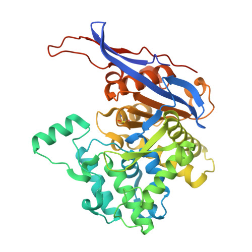
    The ReAV enzyme from Rhizobium etli , a representative of Class 3 L-asparaginases, is sequentially and structurally different from other known L-asparaginases. This distinctiveness makes ReAV a candidate for novel antileukemic therapies. ReAV is a homodimeric protein, with each subunit containing a highly specific zinc-binding site created by two cysteines, a lysine, and a water molecule. Two Ser-Lys tandems (Ser48-Lys51, Ser80-Lys263) are located in the close proximity of the metal binding site, with Ser48 hypothesized to be the catalytic nucleophile. To further investigate the catalytic process of ReAV, site-directed mutagenesis was employed to introduce alanine substitutions at residues from the Ser-Lys tandems and at Arg47, located near the Ser48-Lys51 tandem. These mutational studies, along with enzymatic assays and X-ray structure determinations, demonstrated that substitution of each of these highly conserved residues abolished the catalytic activity, confirming their essential role in enzyme mechanism.


  • Organizational Affiliation
    • Institute of Bioorganic Chemistry, Polish Academy of Sciences, Noskowskiego 12/14, 61-704 Poznan, Poland.

Macromolecules
Find similar proteins by:  (by identity cutoff)  |  3D Structure
Entity ID: 1
MoleculeChains Sequence LengthOrganismDetailsImage
L-asparaginase II
A, B, C, D
373Rhizobium etliMutation(s): 1 
Gene Names: ansA
UniProt
Find proteins for Q9RFN5 (Rhizobium etli)
Explore Q9RFN5 
Go to UniProtKB:  Q9RFN5
Entity Groups  
Sequence Clusters30% Identity50% Identity70% Identity90% Identity95% Identity100% Identity
UniProt GroupQ9RFN5
Sequence Annotations
Expand
  • Reference Sequence
Small Molecules
Ligands 8 Unique
IDChains Name / Formula / InChI Key2D Diagram3D Interactions
PGE
Query on PGE

Download Ideal Coordinates CCD File 
OA [auth C]TRIETHYLENE GLYCOL
C6 H14 O4
ZIBGPFATKBEMQZ-UHFFFAOYSA-N
TRS
Query on TRS

Download Ideal Coordinates CCD File 
GA [auth C],
N [auth A],
T [auth B],
XA [auth D]
2-AMINO-2-HYDROXYMETHYL-PROPANE-1,3-DIOL
C4 H12 N O3
LENZDBCJOHFCAS-UHFFFAOYSA-O
PEG
Query on PEG

Download Ideal Coordinates CCD File 
K [auth A],
NA [auth C],
UA [auth D]
DI(HYDROXYETHYL)ETHER
C4 H10 O3
MTHSVFCYNBDYFN-UHFFFAOYSA-N
SO4
Query on SO4

Download Ideal Coordinates CCD File 
DA [auth C]
EA [auth C]
H [auth A]
L [auth A]
LA [auth C]
DA [auth C],
EA [auth C],
H [auth A],
L [auth A],
LA [auth C],
MA [auth C],
RA [auth D],
S [auth B],
TA [auth D],
V [auth B],
Y [auth B]
SULFATE ION
O4 S
QAOWNCQODCNURD-UHFFFAOYSA-L
GOL
Query on GOL

Download Ideal Coordinates CCD File 
I [auth A],
J [auth A]
GLYCEROL
C3 H8 O3
PEDCQBHIVMGVHV-UHFFFAOYSA-N
ZN
Query on ZN

Download Ideal Coordinates CCD File 
O [auth A],
PA [auth C],
Z [auth B],
ZA [auth D]
ZINC ION
Zn
PTFCDOFLOPIGGS-UHFFFAOYSA-N
EDO
Query on EDO

Download Ideal Coordinates CCD File 
AA [auth C]
BA [auth C]
CA [auth C]
E [auth A]
F [auth A]
AA [auth C],
BA [auth C],
CA [auth C],
E [auth A],
F [auth A],
FA [auth C],
G [auth A],
HA [auth C],
IA [auth C],
JA [auth C],
KA [auth C],
M [auth A],
Q [auth B],
QA [auth D],
R [auth B],
SA [auth D],
U [auth B],
VA [auth D],
W [auth B],
WA [auth D],
X [auth B],
YA [auth D]
1,2-ETHANEDIOL
C2 H6 O2
LYCAIKOWRPUZTN-UHFFFAOYSA-N
CL
Query on CL

Download Ideal Coordinates CCD File 
AB [auth D],
P [auth A]
CHLORIDE ION
Cl
VEXZGXHMUGYJMC-UHFFFAOYSA-M
Modified Residues  1 Unique
IDChains TypeFormula2D DiagramParent
CSO
Query on CSO
A, B, C, D
L-PEPTIDE LINKINGC3 H7 N O3 SCYS
Experimental Data & Validation

Experimental Data

  • Method: X-RAY DIFFRACTION
  • Resolution: 1.60 Å
  • R-Value Free:  0.192 (Depositor), 0.192 (DCC) 
  • R-Value Work:  0.160 (Depositor), 0.161 (DCC) 
  • R-Value Observed: 0.161 (Depositor) 
Space Group: P 1 21 1
Unit Cell:
Length ( Å )Angle ( ˚ )
a = 77.887α = 90
b = 91.507β = 97.08
c = 114.338γ = 90
Software Package:
Software NamePurpose
PHENIXrefinement
XDSdata reduction
XSCALEdata scaling
PHASERphasing

Structure Validation

View Full Validation Report


Entry History & Funding Information

Deposition Data


Funding OrganizationLocationGrant Number
Polish National Science CentrePoland2020/37/B/NZ1/03250

Revision History  (Full details and data files)

  • Version 1.0: 2025-07-16
    Type: Initial release
  • Version 1.1: 2025-08-06
    Changes: Database references