9V69 | pdb_00009v69

X-ray crystal structure of the two-electron reduced form of wild type b5R


Experimental Data Snapshot

  • Method: X-RAY DIFFRACTION
  • Resolution: 0.96 Å
  • R-Value Free: 
    0.148 (Depositor), 0.160 (DCC) 
  • R-Value Work: 
    0.127 (Depositor), 0.144 (DCC) 

Starting Model: experimental
View more details

wwPDB Validation   3D Report Full Report


Ligand Structure Quality Assessment 


This is version 1.0 of the entry. See complete history


Literature

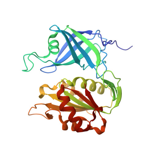
Structural basis of hydride and proton transfer reactions revealed by the detection of hydrogen atoms in mammalian NADH-cytochrome b 5 reductase.

Hirano, Y.Kurihara, K.Kusaka, K.Ostermann, A.Hikita, M.Kimura, S.Miki, K.Tamada, T.

(2025) Structure 

  • DOI: https://doi.org/10.1016/j.str.2025.10.006
  • Primary Citation of Related Structures:  
    9V69, 9V6A, 9V6B, 9V6C

  • PubMed Abstract: 

    Many structural studies have been reported for ferredoxin:NADP + reductase family members, but an experimental validation of the catalytic hydride and proton transfer steps through a direct detection of the involved hydrogen atoms has not been achieved so far. Here, we determined high-resolution X-ray and neutron crystal structures of NADH-cytochrome b 5 reductase, which acts as an electron supplier for various metabolic processes and mediates hydride and proton transfer reactions via its FAD and NADH cofactors. The X-ray structures identify the FADH - -NAD + and FAD-NADH complexes based on the electron densities of the hydrogen atoms bound to the cofactors. The neutron structures determined at different pD-values show a difference in the protonation state of the histidine residue in the hydrogen-bond network from FAD to the protein surface. The observation of the hydrogen atoms reveals the structural basis for the hydride and proton transfer reactions catalyzed by NADH-cytochrome b 5 reductase.


  • Organizational Affiliation
    • Institute for Quantum Life Science, National Institutes for Quantum Science and Technology (QST), Inage, Chiba 263-8555, Japan; PRESTO, The Japan Science and Technology Agency, Kawaguchi 332-0021, Saitama, Japan; Center of Quantum Life Science for Structural Therapeutics (cQUEST), Chiba University, Inage, Chiba 265-8522, Japan. Electronic address: hirano.yu@qst.go.jp.

Macromolecules
Find similar proteins by:  (by identity cutoff)  |  3D Structure
Entity ID: 1
MoleculeChains Sequence LengthOrganismDetailsImage
NADH-cytochrome b5 reductase 3272Sus scrofaMutation(s): 0 
Gene Names: CYB5R3DIA1
EC: 1.6.2.2
UniProt
Find proteins for P83686 (Sus scrofa)
Explore P83686 
Go to UniProtKB:  P83686
Entity Groups  
Sequence Clusters30% Identity50% Identity70% Identity90% Identity95% Identity100% Identity
UniProt GroupP83686
Sequence Annotations
Expand
  • Reference Sequence
Experimental Data & Validation

Experimental Data

  • Method: X-RAY DIFFRACTION
  • Resolution: 0.96 Å
  • R-Value Free:  0.148 (Depositor), 0.160 (DCC) 
  • R-Value Work:  0.127 (Depositor), 0.144 (DCC) 
Space Group: P 21 21 21
Unit Cell:
Length ( Å )Angle ( ˚ )
a = 48.021α = 90
b = 79.255β = 90
c = 86.827γ = 90
Software Package:
Software NamePurpose
SHELXLrefinement
XDSdata reduction
Aimlessdata scaling
PHASERphasing

Structure Validation

View Full Validation Report


Ligand Structure Quality Assessment 


Entry History & Funding Information

Deposition Data


Funding OrganizationLocationGrant Number
Japan Science and TechnologyJapanJPMJPR17G7
Ministry of Education, Culture, Sports, Science and Technology (Japan)JapanPhoton and Quantum Basic Research Coordinated Development Program
Japan Society for the Promotion of Science (JSPS)JapanJP20K06523
Japan Society for the Promotion of Science (JSPS)JapanJP17K07325
Japan Society for the Promotion of Science (JSPS)JapanJP2544037

Revision History  (Full details and data files)

  • Version 1.0: 2025-11-19
    Type: Initial release