9VP7 | pdb_00009vp7

Glucose-6-phosphate dehydrogenase from Leishmania donovani in complex with pseudo substrate


Experimental Data Snapshot

  • Method: X-RAY DIFFRACTION
  • Resolution: 2.25 Å
  • R-Value Free: 
    0.286 (Depositor), 0.290 (DCC) 
  • R-Value Work: 
    0.220 (Depositor), 0.223 (DCC) 

Starting Model: experimental
View more details

wwPDB Validation   3D Report Full Report


Ligand Structure Quality Assessment 


This is version 1.0 of the entry. See complete history


Literature

Crystal structure of leishmanial glucose-6-phosphate dehydrogenase and its implications for selective inhibitor development.

Jakkula, P.Qureshi, I.A.

(2025) Int J Biol Macromol 331: 148313-148313

  • DOI: https://doi.org/10.1016/j.ijbiomac.2025.148313
  • Primary Citation of Related Structures:  
    9VP5, 9VP6, 9VP7

  • PubMed Abstract: 

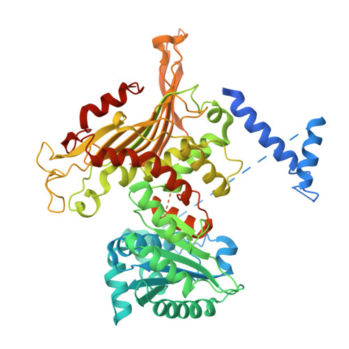
    Glucose-6-phosphate dehydrogenase (G6PDH) is a vital enzyme in trypanosomatid parasites, including Leishmania, where it generates NADPH to combat oxidative stress. In this study, Leishmania donovani G6PDH (LdG6PDH) was cloned, expressed, and purified to homogeneity using chromatographic methods. The purified enzyme existed as a dimer in solution, exhibited optimal activity near physiological pH, and displayed higher affinity for its cofactor (NADP) than for its substrate, glucose-6-phosphate (G6P). The enzymatic activity assay of leishmanial G6PDH revealed that ellagic acid (EA) and epigallocatechin gallate (EGCG) potently inhibit the enzyme, presenting a mixed-type mode of inhibition with respect to the substrate. Fluorescence spectroscopy established that most tryptophan residues are embedded within the hydrophobic core of LdG6PDH, while binding studies confirmed its physical interactions with ligands such as G6P, GLP, NADP, EA, and EGCG. Circular dichroism (CD) spectroscopy divulged a predominance of α-helical structure and substantiated the structural stability of the enzyme under different pH conditions. The crystal structure of LdG6PDH was resolved in both apo and ligand-bound forms, revealing three domains: a unique N-terminal domain, an NADP-binding domain, and a β + α domain. Notably, the ligand-bound structures demonstrated distinct binding positions for G6P and GLP compared to other known G6PDH structures. In silico studies identified lead compounds with a stronger affinity than NADP, indicating favorable physicochemical and pharmacokinetic properties, which were further validated through molecular dynamics simulations and MMPBSA analysis. Collectively, these findings elucidate the structural and functional properties of LdG6PDH, providing a foundation for the development of targeted inhibitors against leishmaniasis.


  • Organizational Affiliation
    • Department of Biotechnology & Bioinformatics, School of Life Sciences, University of Hyderabad, Prof. C.R. Rao Road, Hyderabad, 500046, India.

Macromolecules
Find similar proteins by:  (by identity cutoff)  |  3D Structure
Entity ID: 1
MoleculeChains Sequence LengthOrganismDetailsImage
Glucose-6-phosphate 1-dehydrogenase
A, B
596Leishmania donovaniMutation(s): 0 
Gene Names: g6pdhCGC20_2515CGC21_25385LDBPK_340080LdCL_340005900LDHU3_34.0110
EC: 1.1.1.49
UniProt
Find proteins for A2CIK6 (Leishmania donovani)
Explore A2CIK6 
Go to UniProtKB:  A2CIK6
Entity Groups  
Sequence Clusters30% Identity50% Identity70% Identity90% Identity95% Identity100% Identity
UniProt GroupA2CIK6
Sequence Annotations
Expand
  • Reference Sequence
Small Molecules
Ligands 4 Unique
IDChains Name / Formula / InChI Key2D Diagram3D Interactions
GLP (Subject of Investigation/LOI)
Query on GLP

Download Ideal Coordinates CCD File 
F [auth A]2-amino-2-deoxy-6-O-phosphono-alpha-D-glucopyranose
C6 H14 N O8 P
XHMJOUIAFHJHBW-UKFBFLRUSA-N
PEG
Query on PEG

Download Ideal Coordinates CCD File 
C [auth A],
E [auth A],
G [auth B],
J [auth B]
DI(HYDROXYETHYL)ETHER
C4 H10 O3
MTHSVFCYNBDYFN-UHFFFAOYSA-N
GOL
Query on GOL

Download Ideal Coordinates CCD File 
H [auth B],
I [auth B]
GLYCEROL
C3 H8 O3
PEDCQBHIVMGVHV-UHFFFAOYSA-N
EDO
Query on EDO

Download Ideal Coordinates CCD File 
D [auth A],
K [auth B],
L [auth B],
M [auth B],
N [auth B]
1,2-ETHANEDIOL
C2 H6 O2
LYCAIKOWRPUZTN-UHFFFAOYSA-N
Experimental Data & Validation

Experimental Data

  • Method: X-RAY DIFFRACTION
  • Resolution: 2.25 Å
  • R-Value Free:  0.286 (Depositor), 0.290 (DCC) 
  • R-Value Work:  0.220 (Depositor), 0.223 (DCC) 
Space Group: C 1 2 1
Unit Cell:
Length ( Å )Angle ( ˚ )
a = 191.84α = 90
b = 97.685β = 94.005
c = 70.449γ = 90
Software Package:
Software NamePurpose
REFMACrefinement
CrysalisProdata reduction
CrysalisProdata scaling
MOLREPphasing

Structure Validation

View Full Validation Report


Ligand Structure Quality Assessment 


Entry History & Funding Information

Deposition Data


Funding OrganizationLocationGrant Number
Science and Engineering Research Board (SERB)India--

Revision History  (Full details and data files)

  • Version 1.0: 2025-11-05
    Type: Initial release