21JI | pdb_000021ji

Crystal structure of isoprimeverose-producing enzyme from Phaeoacremonium minimum


Experimental Data Snapshot

  • Method: X-RAY DIFFRACTION
  • Resolution: 2.80 Å
  • R-Value Free: 
    0.268 (Depositor), 0.268 (DCC) 
  • R-Value Work: 
    0.216 (Depositor), 0.216 (DCC) 
  • R-Value Observed: 
    0.218 (Depositor) 

Starting Model: experimental
View more details

wwPDB Validation   3D Report Full Report


Ligand Structure Quality Assessment 


This is version 1.0 of the entry. See complete history


Literature

Structural basis for galactose side-chain recognition in isoprimeverose-producing oligoxyloglucan hydrolase from Phaeoacremonium minimum.

Nakamichi, Y.Watanabe, M.Yaoi, K.Matsuzawa, T.

(2026) Int J Biol Macromol : 151665-151665

  • DOI: https://doi.org/10.1016/j.ijbiomac.2026.151665
  • Primary Citation Related Structures: 
    21JI

  • PubMed Abstract: 

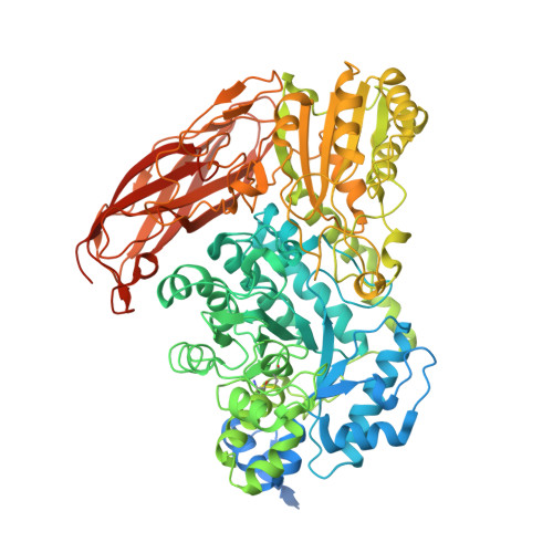
    Isoprimeverose-producing oligoxyloglucan hydrolases (IPases), which belong to glycoside hydrolase family 3 (GH3), release isoprimeverose (α-d-xylopyranosyl-(1→6)-d-glucopyranose) from the non-reducing end of xyloglucan oligosaccharides. The IPase from Phaeoacremonium minimum (PmIPase) exhibits distinctive features compared to its homologous IPase in Aspergillus oryzae, IpeA. The hydrolytic activity of A. oryzae IpeA was inhibited by galactosylation of the second xylopyranosyl residue at the nonreducing end of the substrate, whereas PmIPase was unaffected. To elucidate the structural basis of the unique features of PmIPase, its crystal structure was determined. PmIPase forms a homo-dimer, and its active site is formed by two monomers. Structural comparison with IpeA revealed that the active-site structures are highly conserved between both enzymes, but with some differences, including Phe172 at subsite -1 and Trp624 at a positive subsite in PmIPase (Met173 and Ile625 in IpeA, respectively). Trp624 is from the adjacent monomer and seems to be located at the putative binding position of the galactose residue at subsite +1. When the substrate was a mixture of XXXG, XXLG/XLXG, and XLLG, the W624I mutant showed lower specificity for XLLG than XXXG and XXLG/XLXG, although the wild-type enzyme degraded all three substrates with equal efficiency. This result indicates that Trp624 in PmIPase is involved in the recognition of the galactose residue at subsite +1. These findings provide structural insights into the characteristics and diversity of IPases that adapt to the side chain structures of xyloglucan.


  • Organizational Affiliation
    • Research Institute for Sustainable Chemistry, National Institute of Advanced Industrial Science and Technology (AIST), 3-11-32, Kagamiyama, Higashi-Hiroshima, Hiroshima, 739-0046, Japan.

Macromolecules
Find similar proteins by:  (by identity cutoff)  |  3D Structure
Entity ID: 1
MoleculeChains Sequence LengthOrganismDetailsImage
Isoprimeverose-producing oligoxyloglucan hydrolase,beta-glucosidase
A, B, C
871Rattus rattusPhaeoacremonium minimum
This entity is chimeric
Mutation(s): 0 
Gene Names: RAGUCRPA7_4649
EC: 3.2.1.120 (PDB Primary Data), 3.2.1.21 (PDB Primary Data), 3.2.1.20 (UniProt)
UniProt
Find proteins for A0AA49QB00 (Rattus rattus)
Explore A0AA49QB00 
Go to UniProtKB:  A0AA49QB00
Find proteins for R8BKC2 (Phaeoacremonium minimum (strain UCR-PA7))
Explore R8BKC2 
Go to UniProtKB:  R8BKC2
Entity Groups  
Sequence Clusters30% Identity50% Identity70% Identity90% Identity95% Identity100% Identity
UniProt GroupsA0AA49QB00R8BKC2
Glycosylation
Glycosylation Sites: 8
Sequence Annotations
Expand
  • Reference Sequence
Oligosaccharides

Help

Entity ID: 2
MoleculeChains Length2D Diagram Glycosylation3D Interactions
beta-D-mannopyranose-(1-4)-2-acetamido-2-deoxy-beta-D-glucopyranose-(1-4)-2-acetamido-2-deoxy-beta-D-glucopyranose
D, F, I
3N-Glycosylation
Entity ID: 3
MoleculeChains Length2D Diagram Glycosylation3D Interactions
2-acetamido-2-deoxy-beta-D-glucopyranose-(1-4)-2-acetamido-2-deoxy-beta-D-glucopyranose
E, G, H
2N-Glycosylation
Small Molecules
Ligands 6 Unique
IDChains Name / Formula / InChI Key2D Diagram3D Interactions
P6G
Query on P6G

Download Ideal Coordinates CCD File 
KA [auth B]HEXAETHYLENE GLYCOL
C12 H26 O7
IIRDTKBZINWQAW-UHFFFAOYSA-N
NAG
Query on NAG

Download Ideal Coordinates CCD File 
AA [auth A]
BA [auth A]
CA [auth A]
EA [auth B]
EB [auth C]
AA [auth A],
BA [auth A],
CA [auth A],
EA [auth B],
EB [auth C],
FA [auth B],
FB [auth C],
GB [auth C],
HA [auth B],
J [auth A],
JA [auth B],
K [auth A],
L [auth A],
N [auth A],
O [auth A],
Q [auth A],
UA [auth B],
VA [auth B],
WA [auth B],
XA [auth C],
YA [auth C],
ZA [auth C]
2-acetamido-2-deoxy-beta-D-glucopyranose
C8 H15 N O6
OVRNDRQMDRJTHS-FMDGEEDCSA-N
PG4
Query on PG4

Download Ideal Coordinates CCD File 
P [auth A]TETRAETHYLENE GLYCOL
C8 H18 O5
UWHCKJMYHZGTIT-UHFFFAOYSA-N
PGE
Query on PGE

Download Ideal Coordinates CCD File 
DA [auth A]TRIETHYLENE GLYCOL
C6 H14 O4
ZIBGPFATKBEMQZ-UHFFFAOYSA-N
ACT
Query on ACT

Download Ideal Coordinates CCD File 
BB [auth C]
CB [auth C]
DB [auth C]
GA [auth B]
IA [auth B]
BB [auth C],
CB [auth C],
DB [auth C],
GA [auth B],
IA [auth B],
M [auth A],
MA [auth B],
NA [auth B],
OA [auth B],
PA [auth B],
QA [auth B],
RA [auth B],
S [auth A],
SA [auth B],
T [auth A],
TA [auth B],
U [auth A],
V [auth A],
W [auth A],
X [auth A],
Y [auth A],
Z [auth A]
ACETATE ION
C2 H3 O2
QTBSBXVTEAMEQO-UHFFFAOYSA-M
CA
Query on CA

Download Ideal Coordinates CCD File 
AB [auth C],
LA [auth B],
R [auth A]
CALCIUM ION
Ca
BHPQYMZQTOCNFJ-UHFFFAOYSA-N
Experimental Data & Validation

Experimental Data

  • Method: X-RAY DIFFRACTION
  • Resolution: 2.80 Å
  • R-Value Free:  0.268 (Depositor), 0.268 (DCC) 
  • R-Value Work:  0.216 (Depositor), 0.216 (DCC) 
  • R-Value Observed: 0.218 (Depositor) 
Space Group: C 1 2 1
Unit Cell:
Length ( Å )Angle ( ˚ )
a = 186.185α = 90
b = 74.028β = 95.232
c = 184.494γ = 90
Software Package:
Software NamePurpose
PHENIXrefinement
XDSdata reduction
XDSdata scaling
MOLREPphasing

Structure Validation

View Full Validation Report


Ligand Structure Quality Assessment 


Entry History & Funding Information

Deposition Data


Funding OrganizationLocationGrant Number
Japan Society for the Promotion of Science (JSPS)Japan24K01668

Revision History  (Full details and data files)

  • Version 1.0: 2026-04-08
    Type: Initial release