9O2C | pdb_00009o2c

Heparanase P6 in complex with fragment J72


Experimental Data Snapshot

  • Method: X-RAY DIFFRACTION
  • Resolution: 1.70 Å
  • R-Value Free: 
    0.215 (Depositor), 0.215 (DCC) 
  • R-Value Work: 
    0.194 (Depositor), 0.194 (DCC) 
  • R-Value Observed: 
    0.195 (Depositor) 

Starting Model: experimental
View more details

wwPDB Validation   3D Report Full Report


Ligand Structure Quality Assessment 


This is version 1.1 of the entry. See complete history


Literature

Fragment Screening and Structure-Guided Development of Heparanase Inhibitors Reveal Orthosteric and Allosteric Inhibition.

Davies, L.J.Whitefield, C.Kim, H.Nitsche, C.Jackson, C.J.Frkic, R.L.

(2026) ACS Med Chem Lett 17: 383-390

  • DOI: https://doi.org/10.1021/acsmedchemlett.5c00587
  • Primary Citation of Related Structures:  
    9O1R, 9O1S, 9O1T, 9O1U, 9O1V, 9O1W, 9O1X, 9O1Y, 9O1Z, 9O20, 9O21, 9O22, 9O23, 9O24, 9O25, 9O26, 9O27, 9O28, 9O29, 9O2A, 9O2B, 9O2C, 9O2D, 9O2E, 9O2F, 9O2G, 9O2H, 9O2I, 9O2J, 9O2K, 9O2L, 9O2M

  • PubMed Abstract: 

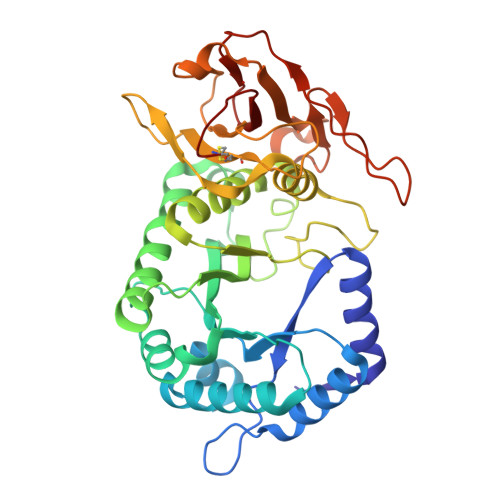
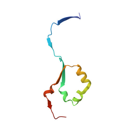
    Heparanase is the sole enzyme responsible for breaking down heparan sulfate within the extracellular matrix, and its overexpression is linked to human diseases. Despite heparanase being a promising drug target, most efforts have focused on substrate mimetics, which have failed clinical trials, highlighting the need for new inhibitor scaffolds. Here, we employed fragment-based drug design to explore a novel chemical space to develop small molecule inhibitors of heparanase. We used a crystallographic and computational approach to identify 31 fragments that bind heparanase; five of these inhibited heparanase in the micromolar range. One of these fragments underwent two cycles of fragment growing, which resulted in a compound with a 7-fold increased potency compared to the initial hit. The results from our fragment screen unveil untapped chemical space for heparanase inhibition, paving the way for the development of potent drug leads with the potential to transform the treatment of heparanase-related diseases.


  • Organizational Affiliation
    • Research School of Chemistry, Australian National University, Canberra, ACT 2601, Australia.

Macromolecules
Find similar proteins by:  (by identity cutoff)  |  3D Structure
Entity ID: 1
MoleculeChains Sequence LengthOrganismDetailsImage
Heparanase 50 kDa subunit382Homo sapiensMutation(s): 0 
Gene Names: HPSEHEPHPAHPA1HPR1HPSE1HSE1
EC: 3.2.1.166
UniProt & NIH Common Fund Data Resources
Find proteins for Q9Y251 (Homo sapiens)
Explore Q9Y251 
Go to UniProtKB:  Q9Y251
PHAROS:  Q9Y251
GTEx:  ENSG00000173083 
Entity Groups  
Sequence Clusters30% Identity50% Identity70% Identity90% Identity95% Identity100% Identity
UniProt GroupQ9Y251
Sequence Annotations
Expand
  • Reference Sequence
Find similar proteins by:  (by identity cutoff)  |  3D Structure
Entity ID: 2
MoleculeChains Sequence LengthOrganismDetailsImage
Heparanase 8 kDa subunit74Homo sapiensMutation(s): 0 
Gene Names: HPSEHEPHPAHPA1HPR1HPSE1HSE1
EC: 3.2.1.166
UniProt & NIH Common Fund Data Resources
Find proteins for Q9Y251 (Homo sapiens)
Explore Q9Y251 
Go to UniProtKB:  Q9Y251
PHAROS:  Q9Y251
GTEx:  ENSG00000173083 
Entity Groups  
Sequence Clusters30% Identity50% Identity70% Identity90% Identity95% Identity100% Identity
UniProt GroupQ9Y251
Sequence Annotations
Expand
  • Reference Sequence
Small Molecules
Ligands 3 Unique
IDChains Name / Formula / InChI Key2D Diagram3D Interactions
GGB (Subject of Investigation/LOI)
Query on GGB

Download Ideal Coordinates CCD File 
G [auth A],
H [auth A]
L-CANAVANINE
C5 H12 N4 O3
FSBIGDSBMBYOPN-VKHMYHEASA-N
SO4
Query on SO4

Download Ideal Coordinates CCD File 
J [auth A],
K [auth A]
SULFATE ION
O4 S
QAOWNCQODCNURD-UHFFFAOYSA-L
EDO
Query on EDO

Download Ideal Coordinates CCD File 
C [auth A],
D [auth A],
E [auth A],
F [auth A],
I [auth A]
1,2-ETHANEDIOL
C2 H6 O2
LYCAIKOWRPUZTN-UHFFFAOYSA-N
Experimental Data & Validation

Experimental Data

  • Method: X-RAY DIFFRACTION
  • Resolution: 1.70 Å
  • R-Value Free:  0.215 (Depositor), 0.215 (DCC) 
  • R-Value Work:  0.194 (Depositor), 0.194 (DCC) 
  • R-Value Observed: 0.195 (Depositor) 
Space Group: P 21 21 21
Unit Cell:
Length ( Å )Angle ( ˚ )
a = 59.311α = 90
b = 75.566β = 90
c = 125.354γ = 90
Software Package:
Software NamePurpose
PHENIXrefinement
Aimlessdata scaling
XDSdata reduction
PHASERphasing

Structure Validation

View Full Validation Report


Ligand Structure Quality Assessment 


Entry History & Funding Information

Deposition Data


Funding OrganizationLocationGrant Number
Australian Research Council (ARC)AustraliaCE200100012
Australian Research Council (ARC)AustraliaCE200100029

Revision History  (Full details and data files)

  • Version 1.0: 2026-02-11
    Type: Initial release
  • Version 1.1: 2026-03-04
    Changes: Database references