


Crystal structure of CD1a presenting ganglioside GD3
Cao, T.P.To be published.
Experimental Data Snapshot
Starting Model: experimental
View more details
Entity ID: 1 | |||||
|---|---|---|---|---|---|
| Molecule | Chains | Sequence Length | Organism | Details | Image |
| T-cell surface glycoprotein CD1a | 285 | Homo sapiens | Mutation(s): 1 Gene Names: CD1A | 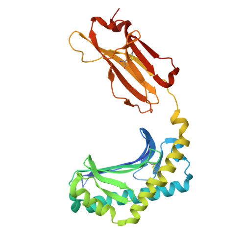 | |
UniProt & NIH Common Fund Data Resources | |||||
Find proteins for P06126 (Homo sapiens) Explore P06126 Go to UniProtKB: P06126 | |||||
PHAROS: P06126 GTEx: ENSG00000158477 | |||||
Entity Groups | |||||
| Sequence Clusters | 30% Identity50% Identity70% Identity90% Identity95% Identity100% Identity | ||||
| UniProt Group | P06126 | ||||
Glycosylation | |||||
| Glycosylation Sites: 1 | Go to GlyGen: P06126-1 | ||||
Sequence AnnotationsExpand | |||||
| |||||
Entity ID: 2 | |||||
|---|---|---|---|---|---|
| Molecule | Chains | Sequence Length | Organism | Details | Image |
| Beta-2-microglobulin | 106 | Homo sapiens | Mutation(s): 0 Gene Names: B2M, CDABP0092, HDCMA22P | 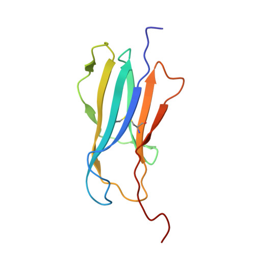 | |
UniProt & NIH Common Fund Data Resources | |||||
Find proteins for P61769 (Homo sapiens) Explore P61769 Go to UniProtKB: P61769 | |||||
PHAROS: P61769 GTEx: ENSG00000166710 | |||||
Entity Groups | |||||
| Sequence Clusters | 30% Identity50% Identity70% Identity90% Identity95% Identity100% Identity | ||||
| UniProt Group | P61769 | ||||
Sequence AnnotationsExpand | |||||
| |||||
| Ligands 2 Unique | |||||
|---|---|---|---|---|---|
| ID | Chains | Name / Formula / InChI Key | 2D Diagram | 3D Interactions | |
| A1CB9 (Subject of Investigation/LOI) Query on A1CB9 | D [auth A] | N-{(2S,3R)-1-[(4-O-beta-D-galactopyranosyl-beta-D-glucopyranosyl)oxy]-3-hydroxyoctadecan-2-yl}tetracosanamide C54 H105 N O13 JRPWXSMBCYEJCN-DJSCMYBVSA-N |  | ||
| NAG Query on NAG | C [auth A] | 2-acetamido-2-deoxy-beta-D-glucopyranose C8 H15 N O6 OVRNDRQMDRJTHS-FMDGEEDCSA-N |  | ||
| Length ( Å ) | Angle ( ˚ ) |
|---|---|
| a = 42.392 | α = 90 |
| b = 90.35 | β = 90 |
| c = 107.445 | γ = 90 |
| Software Name | Purpose |
|---|---|
| PHENIX | refinement |
| PDB_EXTRACT | data extraction |
| XDS | data reduction |
| Aimless | data scaling |
| PHASER | phasing |
| Funding Organization | Location | Grant Number |
|---|---|---|
| Australian Research Council (ARC) | Australia | DE210101031 |


