9PNA | pdb_00009pna

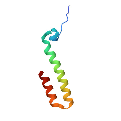
CsgG nanopore in complex with designed CsgX1


Experimental Data Snapshot

  • Method: ELECTRON MICROSCOPY
  • Resolution: 2.61 Å
  • Aggregation State: PARTICLE 
  • Reconstruction Method: SINGLE PARTICLE 

wwPDB Validation 3D Report Full Report

Validation slider image for 9PNA

This is version 1.1 of the entry. See complete history. 

Literature

De novo design of semisynthetic conduction pores.

Schnaider, L., Hatstat, A.K., Scott, A.J., Tan, S.K., Hambley, R.G., Dawson, W.M., Griffiths, R.C., Kormos, R.C., Melo, A.A., Tse, E., Polizzi, N.F., Wallace, E.J., Merz, G.E., DeGrado, W.F.

(2026) Nat Struct Mol Biol 

  • DOI: https://doi.org/10.1038/s41594-026-01881-w
  • Primary Citation Related Structures: 
    9PN8, 9PN9, 9PNA, 9PNB

  • PubMed Abstract: 

    Protein pores used for molecular sensing are generally assembled from large, multi-subunit natural proteins. Their ion conductance, and thus performance in sensing applications, depends on lumen geometry and chemistry. Here rather than designing a new pore or relying only on mutations to a natural scaffold, we show that installing additional de novo subunits to native protein pore complexes enables large-scale architectural changes. We design de novo proteins that integrate seamlessly with CsgG, a pore widely used in sensing applications, to form 18-subunit, 315-kilodalton semisynthetic conduction pores. This required designing within a confined, nonuniform pore under ninefold symmetry and maintaining an open conducting lumen with a stable, low-noise baseline current. The complexes exhibit distinct current-voltage responses relative to the native pore, including clear rectification, while cryo-electron microscopy confirms the designed lumen architecture, establishing a strategy for modifying existing nano-assemblies with designed protein components under stringent symmetry and structural constraints.


  • Organizational Affiliation: 
    • Department of Pharmaceutical Chemistry, University of California, San Francisco, CA, USA.

Macromolecule Content 

  • Total Structure Weight: 325.89 kDa 
  • Atom Count: 21,591 
  • Modeled Residue Count: 2,790 
  • Deposited Residue Count: 2,961 
  • Unique protein chains: 2

Macromolecules

Find similar proteins by:|  3D Structure
Entity ID: 1
MoleculeChains  Sequence LengthOrganismDetailsImage
CsgX1
A, B, C, D, E
A, B, C, D, E, F, G, H, I
57synthetic constructMutation(s): 0 
Entity Groups
Sequence Clusters30% Identity50% Identity70% Identity90% Identity95% Identity100% Identity
Sequence Annotations
Expand
Reference Sequence
Find similar proteins by:|  3D Structure
Entity ID: 2
MoleculeChains  Sequence LengthOrganismDetailsImage
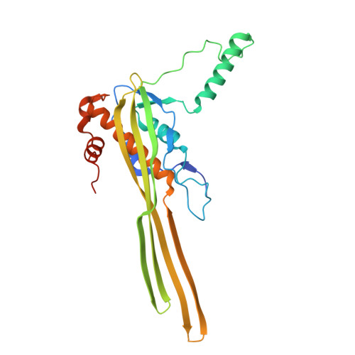
Curli production assembly/transport component CsgG
J, K, L, M, N
J, K, L, M, N, O, P, Q, R
272Escherichia coliMutation(s): 0 
Gene Names: csgG, Z1670, ECs1414
UniProt
Find proteins for P0AEA4 (Escherichia coli O157:H7)
Explore P0AEA4 
Go to UniProtKB:  P0AEA4
Entity Groups
Sequence Clusters30% Identity50% Identity70% Identity90% Identity95% Identity100% Identity
UniProt GroupP0AEA4
Sequence Annotations
Expand
Reference Sequence

Experimental Data & Validation

Experimental Data

  • Method: ELECTRON MICROSCOPY
  • Resolution: 2.61 Å
  • Aggregation State: PARTICLE 
  • Reconstruction Method: SINGLE PARTICLE 
EM Software:
TaskSoftware PackageVersion
RECONSTRUCTIONcryoSPARC

Structure Validation

View Full Validation Report


Entry History 

& Funding Information

Deposition Data


Funding OrganizationLocationGrant Number
National Institutes of Health/National Institute of General Medical Sciences (NIH/NIGMS)United StatesF32GM147962
National Institutes of Health/National Institute of General Medical Sciences (NIH/NIGMS)United StatesK99GM155611

Revision History  (Full details and data files)

  • Version 1.0: 2026-09-16
    Type: Initial release
  • Version 1.1: 2026-10-07
    Changes: Data collection, Database references